
Page 322 - Hematology_ Basic Principles and Practice ( PDFDrive )
P. 322
264 Part III Immunologic Basis of Hematology
factor B by C3(H 2O) allows factor D, another protease, to cleave from membrane surface glycoproteins) found on host surfaces in
factor B to form Ba and Bb. Bb remains associated with C3(H 2O) to contact with blood plasma. Although these polyanion binding sites
form the C3(H 2O)Bb complex. Factor D appears to function as a are not required for FH to regulate fluid phase AP C3 convertase,
serine protease in its native state but can cleave factor B only when they are required for its activity on surface-bound C3bBb. In fact,
bound to C3. Recently, there has been an interesting connection this is the basis for FH being able to discriminate between AP C3
found between factor D and MASP-1, a component of the LP. It was convertase adventitiously deposited on host tissue versus that depos-
found that a MASP-1/MASP-3 knockout mouse (the proteins ited on a microbial surface because the latter do not possess either
MASP-1 and MASP-3 are alternative splice products of the same the sulfated glycosaminoglycans or the sialic acid arrays. 30,31
gene) completely lacked AP functionality. Upon further investigation, Pathogen surfaces are normally not afforded the protection
it was determined that the secreted factor D in this mouse possessed offered by these regulators. Persistence of the C3bBb convertase on
a five-residue propeptide at its amino terminus. Removal of this microbial surfaces may additionally be favored by the positive regula-
propeptide from factor D by the addition of MASP-1 resulted in tor properdin (factor P). This positive modulation of the AP by
restoration of AP functionality. 26 properdin has traditionally been thought to be attributable to its
C3(H 2O)Bb is an enzymatic complex capable of cleaving native ability to prolong the lifetime of the AP C3 convertase by forming a
C3. This complex is a fluid-phase C3 convertase. Although it is C3bBbP complex. This mechanism is still valid, but recently, evi-
formed only in small amounts, it can cleave many molecules of dence has been presented that properdin, which circulates predomi-
C3. Much of the C3b produced in this process is inactivated by nantly as a homotrimer, may also be able to recognize AP targets
hydrolysis, but some attaches covalently to the surface of host cells directly. Specifically, it has been shown to bind to microbial surfaces,
or pathogens. C3b bound in this way is able to bind factor B, such as to Neisseria gonorrhoeae or yeast cell walls, that are known AP
allowing its cleavage by factor D to yield Ba and Bb. The result is activators, but not to strains of Escherichia coli that are known to be
the formation of C3bBb, a C3 convertase akin to C4b2a found in nonactivators of the AP of complement. Because it is a homotrimer,
the classical and MBL pathways, with the capability of initiating an even if factor P uses two of its subunits to bind to the microbial
amplification cascade. surface, one is still left that can recruit C3b, or C3(H 2 O), from the
In light of the nonspecific nature of C3b binding in the AP, it is fluid phase to the microbial surface. The properdin-bound C3b/
not surprising that a number of complement regulators exist both in C3(H2O) can then act as a platform for recruiting factors B and D,
32
the plasma and on host cell membranes to prevent complement thereby forming a surface-bound AP C3 convertase. Consistent
activation on self-tissues. Some of these regulatory components are with this target recognition model for properdin functionality, indi-
mentioned now for the sake of clarity; more detailed attention is viduals with deficiencies in factor P have a heightened susceptibility
provided later in this chapter (Table 24.1). CR1 and DAF (CD55) to infection with Neisseria. 33
compete with factor B for binding to C3b on the cell surface and can After forming, the C3bBb convertase rapidly cleaves more C3
28
displace Bb from a convertase that has already formed. Factor I (FI), to C3b, which can participate in the formation of more molecules
a serum protease, in concert with CR1 or MCP (CD46) can prevent of C3bBb convertase. The AP thereby activates an amplification
convertase formation by converting C3b into its inactive derivative, loop that can proceed on the surface of a pathogen but not on a
29
iC3b. CR1 is unique among the FI cofactors in facilitating an host cell. An additional point regarding amplification by the AP
additional proteolytic cleavage of iC3b to yield C3c and C3dg (see is that C3b deposited on a target as a result of activation of either
Fig. 24.2B). Trimming of the latter by noncomplement proteases the CP or the LP can act as a nidus for the formation of an AP C3
yields the proteolytic limit fragment C3d, which structurally corre- convertase.
sponds to the TED domain (see Fig. 24.2B). Another complement Although specific antibody is not required for AP activation, many
34
regulatory protein found in the plasma is FH. FH binds C3b and is classes of immunoglobulin can facilitate AP activation. The mecha-
able to compete with factor B and displace Bb from the convertase. nism by which this occurs remains elusive, although some evidence
In addition, FH acts as a cofactor for FI to convert C3b to iC3b. In indicates that C3b covalently bound to IgG displays a reduced rate
35
addition to interaction sites for C3b, FH possesses two distinct of inactivation to iC3b by factors H and I. However, in contrast to
binding sites for polyanionic molecules, particularly various sulfated CP activation, which requires Fc, AP activation can occur with
glycosaminoglycans (e.g., heparan sulfate) or arrays of sialic acid (e.g., F(ab)′ 2 fragments.
An instructive demonstration for the role of antibody in continu-
ing the AP cascade, with possible ramifications for human disease,
comes from a murine model of rheumatoid arthritis. Mice do not
36
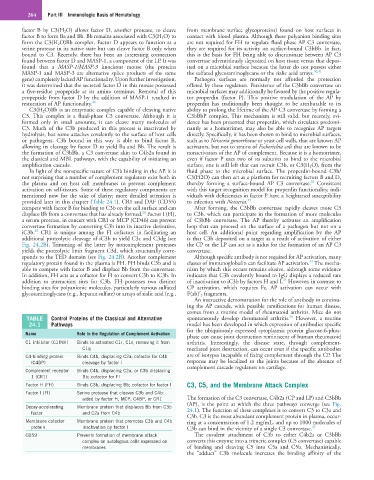
TABLE Control Proteins of the Classical and Alternative spontaneously develop rheumatoid arthritis. However, a murine
24.1 Pathways model has been developed in which expression of antibodies specific
for the ubiquitously expressed cytoplasmic protein glucose-6-phos-
Name Role in the Regulation of Complement Activation
phate can cause joint destruction reminiscent of human rheumatoid
C1 inhibitor (C1INH) Binds to activated C1r, C1s, removing it from arthritis. Interestingly, the disease state, through complement-
C1q mediated joint destruction, can occur even if the specific antibodies
C4-binding protein Binds C4b, displacing C2a; cofactor for C4b are of isotypes incapable of fixing complement through the CP. The
(C4BP) cleavage by factor I response may be localized to the joints because of the absence of
complement cascade regulators on cartilage.
Complement receptor Binds C4b, displacing C2a, or C3b displacing
1 (CR1) Bb; cofactor for FI
Factor H (FH) Binds C3b, displacing Bb; cofactor for factor I C3, C5, and the Membrane Attack Complex
Factor I (FI) Serine protease that cleaves C3b and C4b:
aided by factor H, MCP, C4BP, or CR1 The formation of the C3 convertase, C4b2a (CP and LP) and C3bBb
(AP), is the point at which the three pathways converge (see Fig.
Decay-accelerating Membrane protein that displaces Bb from C3b 24.1). The function of these complexes is to convert C3 to C3a and
factor and C2a from C4b
C3b. C3 is the most abundant complement protein in plasma, occur-
Membrane cofactor Membrane protein that promotes C3b and C4b ring at a concentration of 1.2 mg/mL, and up to 1000 molecules of
protein inactivation by factor I C3b can bind in the vicinity of a single C3 convertase. 37
CD59 Prevents formation of membrane attack The covalent attachment of C3b to either C4b2a or C3bBb
complex on autologous cells expressed on converts this enzyme into a trimeric complex (C5 convertase) capable
membranes of binding and cleaving C5 into C5a and C5b. Mechanistically,
the “adduct” C3b molecule increases the binding affinity of the