
Page 423 - Hematology_ Basic Principles and Practice ( PDFDrive )
P. 423
344 Part IV Disorders of Hematopoietic Cell Development
mutation (GATA1 V205M ) significantly impairs FOG-1 binding, but ATG ATG
retains normal DNA affinity based on electromobility shift assays
using synthetic oligonucleotides. This is consistent with the location
of this residue on the surface of the zinc finger opposite the DNA AD N C GATA-1
binding face.
Several other GATA1 mutations have been linked to cases of 1
familial X-linked macrothrombocytopenia with or without anemia.
These substitutions all impair FOG-1 binding, although to different N C GATA-1s
degrees. Substitution of glycine by serine at codon 208 (GATA1 G208S )
results in moderate to severe thrombocytopenia and mild dyseryth- 84 414
ropoiesis, but no anemia. Substitution of the same residue by arginine
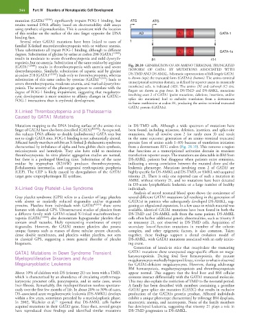
(GATA1 G208R ) results in thrombocytopenia with anemia and severe Fig. 28.10 GENERATION OF AN AMINO TERMINAL TRUNCATED
dyserythropoiesis. Similarly, substitution of aspartic acid by glycine ISOFORM OF GATA1 BY MUTATIONS ASSOCIATED WITH
at codon 218 (GATA1 D218G ) leads only to thrombocytopenia, whereas DS-TMD AND DS-AMKL. Schematic representation of full-length GATA1
substitution of this same codon by tyrosine (GATA1 D218Y ) leads to is shown (top); the truncated form (GATA1s) (bottom). The amino terminal
severe thrombocytopenia, moderate anemia, and marked dyserythro- transcriptional activation domain, as defined by reporter assays in transiently
poiesis. The severity of the phenotype appears to correlate with the transfected cells, is indicated (AD). The amino (N) and carboxyl (C) zinc
degree of FOG-1 binding impairment, suggesting that megakaryo- fingers are shown as gray boxes. In DS-TMD and DS-AMKL, mutations
cytic development is more sensitive to affinity changes in GATA1– involving exon 2 of GATA1 (point mutations, deletions, insertions, and/or
FOG-1 interactions than is erythroid development. splice site mutations) lead to exclusive translation from a downstream
in-frame methionine at codon 84, producing the amino terminal truncated
GATA1 protein (GATA1s).
X-Linked Thrombocytopenia and β-Thalassemia
Caused by GATA1 Mutations
Mutations mapping to the DNA binding surface of the amino zinc in DS-TMD cells. Although a wide spectrum of mutations have
finger of GATA1 have also been described (GATA1 R216Q ). As expected, been found, including missense, deletion, insertion, and splice-site
this reduces DNA affinity to double (palindromic) GATA sites but mutations, they all involve exon 2 (or rarely exon 3) and result
not to single GATA sites. FOG-1 binding is not substantially altered. in the same outcome: generation of an amino terminal truncated
Affected family members exhibit an X-linked β-thalassemia syndrome protein (loss of amino acids 1−83) because of translation initiation
characterized by imbalance of alpha and beta globin chain synthesis, from a downstream ATG codon (Fig. 28.10). This removes a region
reticulocytosis and hemolysis. They also have mild to moderate that functions as a transcriptional activation domain in transient
thrombocytopenia. In vitro platelet aggregation studies are normal, transfection reporter assays. The mutations are detectable in BM from
but there is a prolonged bleeding time. Substitution of the same DS-AMKL patients but disappear when patients enter remission,
residue by tryptophan (R216W) produces thrombocytopenia, indicating a strong correlation between the mutated clone and the
β-thalassemia intermedia, and congenital erythropoietic porphyria leukemic phenotype. Mutations involving exon 2 of GATA1 are
(CEP). The CEP is likely caused by dysregulation of the GATA1 highly specific for DS-AMKL and DS-TMD, or AMKL with acquired
target gene uroporphyrinogen III synthase. trisomy 21. There is only one reported case of such a mutation in
AMKL without trisomy 21, and no mutations have been detected
in DS-acute lymphoblastic leukemia or a large number of healthy
X-Linked Gray Platelet–Like Syndrome individuals.
Analysis of stored neonatal blood spots shows the coexistence of
Gray platelet syndrome (GPS) refers to a disorder of large platelets several different GATA1 mutations (all resulting in the generation of
with absent or markedly reduced α-granules and/or α-granule GATA1s) in patients who subsequently developed DS-AMKL, sug-
proteins. Platelets from individuals with GATA1 R261Q share some gesting an oligoclonal expansion. In a few cases in which material was
features with classical GPS. Ultrastructural studies of platelets from available, identical GATA1 mutations have been found in both the
a different family with GATA1-related X-linked macrothrombocy- DS-TMD and DS-AMKL cells from the same patient. DS-AMKL
topenia (GATA1 G208S ) also demonstrate hypogranular platelets that cells often harbor additional genetic abnormalities, such as trisomy 8
contain small vacuoles, likely representing membranes of empty or tetrasomy 21, not observed in DS-TMD cells. Acquisition of
α-granules. However, the GATA1 mutant platelets also possess secondary loss-of-function mutations in member of the cohesin
unique features such as masses of dense tubular system channels, complex, and other epigenetic factors, is also common. Taken
dense double membranes, and platelets within platelets, not seen together, these findings support a clonal evolution model of
in classical GPS, suggesting a more general disorder of platelet DS-AMKL, with GATA1 mutations associated with an early initiat-
biogenesis. ing event.
Generation of knock-in mice that recapitulate the truncating
GATA1 Mutations in Down Syndrome Transient GATA1 mutations show unexpected stage-specific effects on mega-
Myeloproliferative Disorders and Acute karyocytopoiesis. During fetal liver hematopoiesis, the mutant
megakaryocytes markedly hyperproliferate, similar to what is observed
Megakaryoblastic Leukemia for GATA1-deficient megakaryocytes. However, during adult-stage
BM hematopoiesis, megakaryocytopoiesis and thrombocytopoiesis
About 10% of children with DS (trisomy 21) are born with a TMD, appear normal. This suggests that the fetal liver and BM cellular
which is characterized by an abundance of circulating erythromega- contexts interact differentially with the GATA1 truncated molecule.
karyocytic precursor cells, pancytopenia, and in some cases, severe This may also explain the restriction of TMD to the neonatal period.
liver fibrosis. Remarkably, this myeloproliferation resolves spontane- A family has been described with members containing a germline
ously over the first few months of life. In about 20% to 30% of cases, GATA1 gene splice site mutation (G332C) that results in exclusive
DS–associated acute megakaryocytic leukemia (DS-AMKL) develops production of the GATA1s protein product. Affected individuals
within a few years, sometimes preceded by a myelodysplastic phase. exhibit a unique phenotype characterized by trilineage BM dysplasia,
23
In 2002, Wechsler et al. reported that DS-AMKL cells harbor macrocytic anemia, and neutropenia. None of the family members
acquired mutations in their GATA1 gene. Since then, several groups has developed leukemia, suggesting that trisomy 21 plays a role in
have reproduced these findings and identified similar mutations DS-TMD progression to DS-AMKL.