
Page 462 - Hematology_ Basic Principles and Practice ( PDFDrive )
P. 462
Chapter 29 Inherited Bone Marrow Failure Syndromes 383
contrast to the other two forms, CDA III is inherited as an autosomal
dominant disorder. The responsible gene is KIF23, which encodes
mitotic kinesin-like protein 1 (MKLP1) that localizes to the midbody
ring. In vitro studies showed that the mutations in the genes result
in cytokinesis failure. It has also been shown that ubiquitination of
MKLP1 is required for midbody ring degradation by autophagy;
thus abrogation of this process may cause cytokinesis failure and
multinuclearity.
In a Swedish family with 31 cases inherited in an autosomal
dominant mode, an excess number of cases with a monoclonal gam-
mopathy and myeloma have occurred. Also, an adult patient with
CDA III was described with T-cell non-Hodgkin lymphoma. These
cases, plus a case of Hodgkin disease occurring in an additional
patient, may indicate an increased incidence of lymphoproliferative
diseases in CDA III.
Laboratory Abnormalities. In CDA III, splenomegaly is usually
minimal or absent. The anemia is usually mild to moderate, but
transfusion-dependent patients have been observed. The circulating
RBCs can be normal or mildly macrocytic. BM examination shows
erythroid hyperplasia. Giant erythroblasts with up to 12 nuclei are
the most distinctive feature of CDA III observed on light microscopic
examination of the BM. These may appear similar to some of the
large multinucleated cells seen in CDA II (see Fig. 29.14). Abnor-
mally large lobulated nuclei and discordance in nuclear maturation
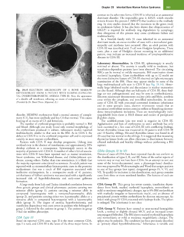
Fig. 29.15 ELECTRON MICROSCOPY OF A BONE MARROW are also found. Although they are hallmarks of CDA III, these find-
ERYTHROBLAST FROM A PATIENT WITH HEMPAS (CONGENI- ings are not pathognomonic and may be seen in erythroleukemia.
TAL DYSERYTHROPOIETIC ANEMIA TYPE II). Note the appearance Electron microscopy demonstrates nuclear clefts and blebs, autolytic
of a double cell membrane, reflecting an excess of endoplasmic reticulum. areas within the cytoplasm, and iron-filled mitochondria. In some
(Provided by Dr. Raoul Fresco, Maywood, IL.) cases of CDA III with presumed autosomal dominant inheritance
and in some sporadic cases, electron microscopy reveals that an
occasional erythroblast section contains stellate or branching electron-
dense intracytoplasmic inclusions. These are morphologically indis-
disorder. HEMPAS erythrocytes bind a normal amount of comple- tinguishable from those in HbH disease and consist of precipitated
ment (C1), but more antibody and less C4 than normal. This causes β-globin chains.
binding of an excess of C3 and hemolysis. The acidified-serum lysis test result is negative in CDA III.
The number of erythroid progenitors is probably normal in BM Agglutination and lysis of erythrocytes to anti-i antibody has only
and blood. Although one study found only normal morphology of been examined in a few cases of CDA III with conflicting findings.
the erythroblasts produced in culture, subsequent studies reported Serum thymidine kinase was measured in 20 patients with CDA III
multinuclearity similar to that seen in the BM. As in CDA I, the and 10 healthy siblings. Elevated thymidine kinase was found in all
defect in CDA II is in the erythroid progenitor cell and is expressed 20 cases but was normal in the siblings. It is suggested that measuring
variably in more mature erythroblasts. thymidine kinase levels can allow clinicians to discriminate between
Patients with CDA II may develop progressive, lifelong iron affected individuals and healthy siblings without performing a BM
overload even in the absence of transfusions, and approximately 20% aspirate.
develop cirrhosis as a consequence. Splenomegaly occurs in the
majority of patients with CDA II. A number of other clinical associa- CDAs Groups IV to VII
tions with CDA II have been reported such as mental retardation, Dozens of cases of CDA have been reported that do not conform to
Sweet syndrome, von Willebrand disease, and Dubin-Johnson syn- the classification of types I, II, and III. Some of the earlier reports of
drome, among others. Rather than true associations, it is likely that variants may or may not have been CDAs. In an attempt to sort out
the majority represent coincidental occurrences. An adult patient was some of the better-documented cases, a phenotype-based classifica-
reported with an extramedullary hematopoietic mass in the posterior tion was proposed by Wickramasinghe and Wood that assigns patients
mediastinum that was a result of BM expansion associated with to one of four groups (not types), designated groups IV, V, VI, and
ineffective erythropoiesis. In a retrospective study of 41 patients, VII. To qualify for inclusion in this classification, each group contains
coinheritance of Gilbert syndrome was associated with a significantly cases from three or more unrelated families. The features of each are
increased risk of hyperbilirubinemia and early-onset gallstone as follows.
formation.
A large study from Italy and Turkey identified correlation between CDA Group IV. This group has severe anemia, transfusion depen-
three genetic groups and clinical phenotype: patients carrying two dence from birth, marked erythroid hyperplasia, normoblastic or
missense alleles (group 1), patients carrying a missense allele in mild to moderate megaloblastic changes, up to 8% BM erythroblasts
compound heterozygosity with a nonsense/hypomorphic allele with markedly irregular or karyorrhectic nuclei, and an absence of
(group 2), and patients carrying either two hypomorphic alleles or a precipitated protein within erythroblasts by electron microscopy. An
nonsense allele in compound heterozygosity with a hypomorphic infant with group IV CDA presented with hydrops fetalis. The spleen
allele (group 3). The degree of anemia, hyperferritenemia, and is enlarged. The inheritance is not clear.
transfusion dependency was most severe in patients who belonged to
group 2, followed by the patients in group 1. The patients in group CDA Group V. Patients have normal or near-normal hemoglobin
3 had the mildest phenotype. levels, a normal or slightly elevated MCV, and an increased serum
unconjugated bilirubin. The BM shows marked erythroid hyperplasia
CDA Type III and normoblastic or mild to moderate megaloblastic changes. The
Based on reported CDA cases, type II is the most common CDA, spleen may be palpable. The condition has been previously described
type I is next, and CDA III is the rarest of the three major forms. In as “primary shunt hyperbilirubinemia.” Inheritance is variable and