
Page 567 - Hematology_ Basic Principles and Practice ( PDFDrive )
P. 567
482 Part V Red Blood Cells
iron therapy. Iron deficiency has other nonhematologic consequences, restricted, frank iron-deficiency anemia develops (see box on Plasma
including impaired immunity and resistance to infection, diminished Iron Concentration and Transferrin Saturation).
exercise tolerance and work performance, and a variety of behavioral Chronic, long-standing iron-deficiency anemia may produce
and neuropsychologic abnormalities. In patients with iron deficiency severe microcytosis and hypochromia, with very pale, distorted RBCs
and heart failure, clinical trials have provided evidence that treatment and dramatic reductions in the mean corpuscular volume and mean
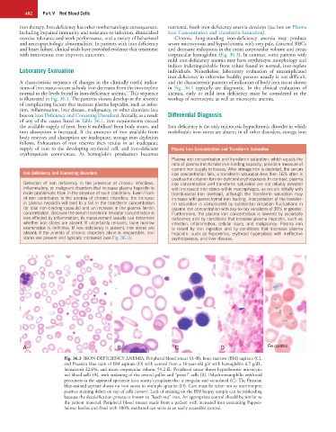
with intravenous iron improves outcomes. corpuscular hemoglobin (Fig. 36.3). In contrast, some patients with
mild iron-deficiency anemia may have erythrocyte morphology and
indices indistinguishable from values found in normal, iron-replete
Laboratory Evaluation individuals. Nonetheless, laboratory evaluation of uncomplicated
iron deficiency in otherwise healthy persons usually is not difficult,
A characteristic sequence of changes in the clinically useful indica- and the characteristic patterns of indicators of body iron status shown
tions of iron status occurs as body iron decreases from the iron-replete in Fig. 36.1 typically are diagnostic. In the clinical evaluation of
6
normal to the levels found in iron-deficiency anemia. This sequence anemia, early or mild iron deficiency must be considered in the
is illustrated in Fig. 36.1. The patterns shown develop in the absence workup of normocytic as well as microcytic anemia.
of complicating factors that increase plasma hepcidin, such as infec-
tion, inflammation, liver disease, malignancy, or other disorders (see
box on Iron Deficiency and Coexisting Disorders). Initially, as a result Differential Diagnosis
of any of the causes listed in Table 36.1, iron requirements exceed
the available supply of iron. Iron is mobilized from body stores, and Iron deficiency is the only microcytic hypochromic disorder in which
iron absorption is increased. If the amounts of iron available from mobilizable iron stores are absent; in all other disorders, storage iron
body reserves and absorption are inadequate, storage iron depletion
follows. Exhaustion of iron reserves then results in an inadequate
supply of iron to the developing erythroid cell, and iron-deficient Plasma Iron Concentration and Transferrin Saturation
erythropoiesis commences. As hemoglobin production becomes
Plasma iron concentration and transferrin saturation, which equals the
ratio of plasma iron to total iron-binding capacity, provide a measure of
current iron supply to tissues. After storage iron is depleted, the serum
Iron Deficiency and Coexisting Disorders iron concentration falls; a transferrin saturation less than 16% often is
used as the criterion for iron-deficient erythropoiesis. In contrast, plasma
Detection of iron deficiency in the presence of chronic infectious, iron concentration and transferrin saturation are not reliably elevated
inflammatory, or malignant disorders that increase plasma hepcidin is with increased iron stores within macrophages, as occurs initially with
more problematic than in the absence of such conditions. Even if lack transfusional iron overload, although the transferrin saturation may
of iron contributes to the anemia of chronic disorders, the increase increase with parenchymal iron loading. Interpretation of the transfer-
in plasma hepcidin will lead to a fall in the transferrin concentration rin saturation is complicated by substantial circadian fluctuations in
(or total iron-binding capacity) and an increase in the plasma ferritin plasma iron concentration with day-to-day variations of 30% or greater.
concentration. Because the serum transferrin receptor concentration is Furthermore, the plasma iron concentration is lowered by ascorbate
less affected by inflammation, its measurement usually can determine deficiency and by conditions that increase plasma hepcidin, such as
whether iron stores are absent. If uncertainty remains, bone marrow infection, inflammation, cellular injury, and malignancy. Plasma iron
examination is definitive. If iron deficiency is present, iron stores are is raised by iron ingestion and by conditions that decrease plasma
absent; if the anemia of chronic disorders alone is responsible, iron hepcidin, such as hypoxemia, erythroid hyperplasia with ineffective
stores are present and typically increased (see Fig. 36.1). erythropoiesis, and liver disease.
A B C D Fe control
Fig. 36.3 IRON-DEFICIENCY ANEMIA. Peripheral blood smear (A–B), bone marrow (BM) aspirate (C),
and Prussian blue stain of BM aspirate (D) with control from a 16-year-old girl with hemoglobin 6.7 g/dL,
hematocrit 22.6%, and mean corpuscular volume 59.2 fL. Peripheral smear shows hypochromic microcytic
red blood cells (A), with widening of the central pallor and “pencil” cells (B). Polychromatophilic erythroid
precursors in the aspirated specimen have scanty cytoplasm that is irregular and vacuolated (C). The Prussian
blue-stained aspirate shows no iron stores in multiple spicules (D). Care must be taken not to overinterpret
positive staining debris on top of cells (center). Lack of staining on the BM biopsy sample can be misleading
because the decalcification process is known to “leach out” iron. An appropriate control should be similar to
the patient material. Peripheral blood smears made from a patient with increased iron-containing Pappen-
heimer bodies and fixed with 100% methanol can serve as an easily accessible control.