
Page 60 - Hematology_ Basic Principles and Practice ( PDFDrive )
P. 60
32 Part I Molecular and Cellular Basis of Hematology
expertise, and instrumentation as well as a large amount of starting are not suitable for the analysis of large numbers of proteins, because
material, all of which tended to make systematic proteomic experi- each protein to be interrogated requires a separate slide. Nevertheless,
ments difficult to perform routinely. However, newer instruments RPPA remains a useful tool in the armamentarium of proteomic
and methods allow for the analysis of significantly more complex research and may prove particularly useful for the comparison of
mixtures, with increased sensitivity and speed. Sequence assignment proteins of interest across a large panel of samples (e.g., across a
confidence, especially for modified peptides, has also been markedly collection of patient samples or cell lines).
improved owing to the increase in both resolution and mass accuracy.
For example, in mammalian cells, it is possible to confidently detect
more than 8000 unique proteins and more than 15,000 phospho- Bead-Based Profiling
peptides in a few days using a single instrument. Efforts to perform
proteome-wide analysis of complex samples such as cells and tissues Another proteomic method involves the multiplexed analysis of
without extensive fractionation represent one end of the spectrum; protein abundance or phosphorylation. Phosphorylation involves the
proteomic analysis with single-cell resolution represents the other use of microspheres (beads). In this approach, a different protein-
end. Although these methods (single-cell Western blot analysis and specific antibody is coupled to beads of distinct color. A mixture of
flow cytometry for intracellular proteins) can currently interrogate antibody-coupled beads is then mixed with protein lysate, and then
only one or a few analytes at the same time, these methods will evolve binding events are detected with a labeled secondary antibody (e.g.,
with ongoing technological progress. antiphosphotyrosine antibody). Multiple analytes are thereby simul-
taneously profiled in a single sample. This approach was successfully
used to profile the tyrosine phosphorylation status of nearly all
Reverse Phase Lysates protein tyrosine kinases across a panel of cell lines. The advantage of
this approach is that multiple proteins (as many as 100 or more) can
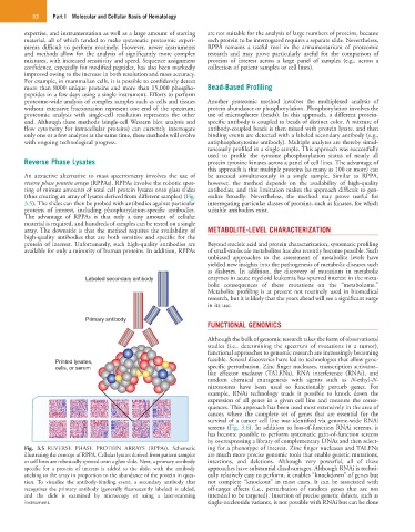
An attractive alternative to mass spectrometry involves the use of be assessed simultaneously in a single sample. Similar to RPPA,
reverse phase protein arrays (RPPAs). RPPAs involve the robotic spot- however, the method depends on the availability of high-quality
ting of minute amounts of total cell protein lysates onto glass slides antibodies, and this limitation makes the approach difficult to gen-
(thus creating an array of lysates derived from different samples) (Fig. eralize broadly. Nevertheless, the method may prove useful for
3.5). The slides can then be probed with antibodies against particular interrogating particular classes of proteins, such as kinases, for which
proteins of interest, including phosphorylation-specific antibodies. suitable antibodies exist.
The advantage of RPPAs is that only a tiny amount of cellular
material is required, and hundreds of samples can be tested on a single
array. The downside is that the method requires the availability of METABOLITE-LEVEL CHARACTERIZATION
high-quality antibodies that are both sensitive and specific for the
protein of interest. Unfortunately, such high-quality antibodies are Beyond nucleic acid and protein characterization, systematic profiling
available for only a minority of human proteins. In addition, RPPAs of small-molecule metabolites has also recently become possible. Such
unbiased approaches to the assessment of metabolite levels have
yielded new insights into the pathogenesis of metabolic diseases such
as diabetes. In addition, the discovery of mutations in metabolic
Labeled secondary antibody enzymes in acute myeloid leukemia has spurred interest in the meta-
bolic consequences of these mutations on the “metabolome.”
Metabolite profiling is at present not routinely used in biomedical
research, but it is likely that the years ahead will see a significant surge
in its use.
Primary antibody
FUNCTIONAL GENOMICS
Although the bulk of genomic research takes the form of observational
studies (i.e., determining the spectrum of mutations in a tumor),
functional approaches to genomic research are increasingly becoming
Printed lysates, feasible. Several discoveries have led to technologies that allow gene-
cells, or serum specific perturbation. Zinc finger nucleases, transcription activator–
like effector nucleases (TALENs), RNA interference (RNAi), and
random chemical mutagenesis with agents such as N-ethyl-N-
nitrosourea have been used to functionally perturb genes. For
example, RNAi technology made it possible to knock down the
expression of all genes in a given cell line and measure the conse-
quences. This approach has been used most extensively in the area of
cancer, where the complete set of genes that are essential for the
survival of a cancer cell line was identified via genome-wide RNAi
screens (Fig. 3.6). In addition to loss-of-function RNAi screens, it
has become possible to perform systematic gain-of-function screens
by overexpressing a library of complementary DNAs and then select-
Fig. 3.5 REVERSE PHASE PROTEIN ARRAYS (RPPAs). Schematic ing for a phenotype of interest. Zinc finger nucleases and TALENs
illustrating the concept of RPPA. Cellular lysates derived from patient samples are much more precise genomic tools that enable genetic mutations,
or cell lines are robotically spotted onto a glass slide. Next, a primary antibody insertions, and deletions. Although very powerful, all of these
specific for a protein of interest is added to the slide, with the antibody approaches have substantial disadvantages. Although RNAi is techni-
sticking to the array in proportion to the abundance of the protein in ques- cally relatively easy to perform, it enables “knockdown” of genes but
tion. To visualize the antibody-binding event, a secondary antibody that not complete “knockout” in most cases. It can be associated with
recognizes the primary antibody (generally fluorescently labeled) is added, off-target effects (i.e., perturbation of random genes that are not
and the slide is examined by microscopy or using a laser-scanning intended to be targeted). Insertion of precise genetic defects, such as
instrument. single-nucleotide variants, is not possible with RNAi but can be done