
Page 622 - Hematology_ Basic Principles and Practice ( PDFDrive )
P. 622
Chapter 39 Megaloblastic Anemias 531
Cobalamin deficiency eventually develops in 10% to 20% of thyroiditis (11%), vitiligo (8%), Addison disease, idiopathic hypo-
patients 8 years after partial gastrectomy; a minority (about 5%) parathyroidism, primary ovarian failure, myasthenia gravis, type 1
develops frank clinical manifestations of cobalamin deficiency with diabetes mellitus, and adult hypogammaglobulinemia. 15,22
megaloblastic anemia. The cause is multifactorial, and contributing Autoimmune gastritis progresses over decades to atrophic body
factors include decreased IF secretion, hypochlorhydria, intestinal gastritis and pernicious anemia. Serum anti-IF antibodies are highly
bacterial overgrowth of cobalamin-consuming organisms, and associ- specific (100%) for pernicious anemia, but the sensitivity is only
ated iron deficiency. The degree of cobalamin deficiency depends on about 50%. Earlier, the clinical use of antiparietal cell antibodies was
the size of the remaining gastric remnant. It is more common in limited because of low specificity. This necessitated use of additional
Bilroth II than in Bilroth I surgery, and in subtotal than in partial surrogate markers (high serum gastrin and low pepsinogen I levels)
gastrectomy. Morbidly obese patients treated surgically with gastric that reflected loss of acid- and IF-secreting parietal (oxyntic) cells.
bypass also have more food-cobalamin malabsorption than patients However, newer enzyme-linked immunosorbent assays (ELISA) for
+
+
22
treated with vertical banded gastroplasty. Even after laparoscopic antiparietal cell antibodies, which are directed against gastric H /K
Roux-en-Y gastric bypass, and despite multivitamin supplementa- ATPase, are 30% more sensitive than previous (immunofluorescence)
tion, iron deficiency was seen in one-half of patients and cobalamin assays. A reanalysis of the clinical utility of combining anti-IF and
166
deficiency seen in one-quarter at 3 years ; therefore these patients newer antiparietal cell antibody tests to noninvasively diagnose perni-
169
probably need higher oral cobalamin (or addition of parenteral) cious anemia points to this approach as very promising. Thus
therapy. among 81 patients with biopsy-proven atrophic body gastritis and
pernicious anemia, combining anti-IF antibodies (37% sensitivity;
Absent Intrinsic Factor Secretion and 100% specificity) with newer antiparietal cell antibodies (sensitivity
91%; specificity 90%) significantly increased their diagnostic perfor-
Pernicious Anemia mance for pernicious anemia, yielding overall 73% sensitivity while
maintaining 100% specificity. 169
A common cause of cobalamin malabsorption is pernicious anemia, Juvenile pernicious anemia can manifest in the second decade with
an autoimmune disease in which the fundamental defect is atrophy severe cobalamin deficiency in conjunction with many of the associ-
of the gastric (parietal cell) oxyntic mucosa that eventually leads to ated endocrinopathies and autoantibodies observed in adults. 15
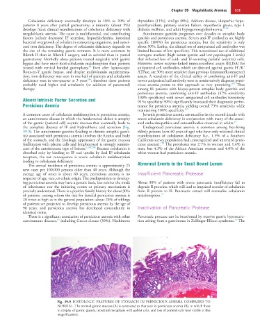
the complete absence of IF and hydrochloric acid secretion (Fig. Undiagnosed pernicious anemia is common among free-living
39.9). The autoimmune gastritis (leading to chronic atrophic gastri- elderly persons (over 60 years of age) who have only minimal clinical
tis) associated with pernicious anemia involves the fundus and body manifestations of cobalamin deficiency (i.e., 1.9% of a Southern
of the stomach, and the histologic appearance of the gastric mucosa California survey population had unrecognized and untreated perni-
170
(infiltration with plasma cells and lymphocytes) is strongly reminis- cious anemia). The prevalence was 2.7% in women and 1.4% in
cent of the autoimmune type of lesions. 6,167,168 Because cobalamin is men, but 4.3% of the African American women and 4.0% of the
absorbed only by binding to IF and uptake by ileal IF-cobalamin white women had pernicious anemia.
receptors, the net consequence is severe cobalamin malabsorption
leading to cobalamin deficiency.
The annual incidence of pernicious anemia is approximately 25 Abnormal Events in the Small Bowel Lumen
new cases per 100,000 persons older than 40 years. Although the
average age of onset is about 60 years, pernicious anemia is no Insufficient Pancreatic Protease
respecter of age, race, or ethnic origin. The predisposition to develop-
ing pernicious anemia may have a genetic basis, but neither the mode About 30% of patients with severe pancreatic insufficiency fail to
of inheritance nor the initiating events or primary mechanism is degrade R proteins, which will lead to impaired transfer of cobalamin
precisely understood. There is a positive family history for about 30% from R protein to IF. Pancreatic extract will normalize cobalamin
of patients, among whom the risk for familial pernicious anemia is malabsorption. 15
20 times as high as in the general population; about 20% of siblings
of patients are projected to develop pernicious anemia by the age of
90 years, and pernicious anemia has developed concordantly in Inactivation of Pancreatic Protease
identical twins.
There is a significant association of pernicious anemia with other Pancreatic protease can be inactivated by massive gastric hypersecre-
15
15
autoimmune diseases, including Graves disease (30%), Hashimoto tion arising from a gastrinoma in Zollinger-Ellison syndrome. The
A B
Fig. 39.9 HISTOLOGIC FEATURES OF STOMACH IN PERNICIOUS ANEMIA COMPARED TO
NORMAL. The normal gastric mucosa (A) is contrasted to that seen in pernicious anemia (B), in which there
is atrophy of gastric glands, intestinal metaplasia with goblet cells, and loss of parietal cells (not visible at this
magnification).