
Page 731 - Hematology_ Basic Principles and Practice ( PDFDrive )
P. 731
618 Part V Red Blood Cells
Cysteine Glutamic acid
ATP
γ−Glutamylcysteine
synthetase ADP
5-Oxoproline γ−Glutamylcysteine
Glycine
Glutathione ATP
synthetase
ADP
H2O2 H2O
Glutathione peroxidase
GSH GSSG
Glutathione reductase
NADP NADPH
G6PD
G6P Pentose F6P
shunt
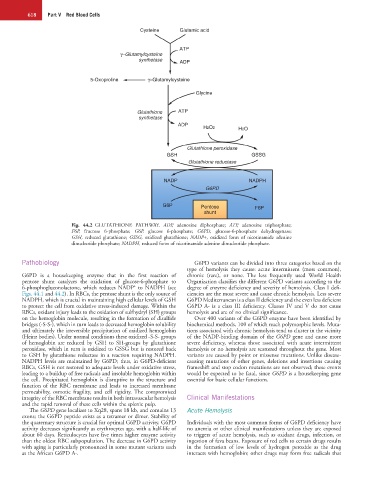
Fig. 44.2 GLUTATHIONE PATHWAY. ADP, adenosine diphosphate; ATP, adenosine triphosphate;
F6P, fructose 6-phosphate; G6P, glucose 6-phosphate; G6PD, glucose-6-phosphate dehydrogenase;
GSH, reduced glutathione; GSSG, oxidized glutathione; NADP+, oxidized form of nicotinamide adenine
dinucleotide phosphate; NADPH, reduced form of nicotinamide adenine dinucleotide phosphate.
Pathobiology G6PD variants can be divided into three categories based on the
type of hemolysis they cause: acute intermittent (most common),
G6PD is a housekeeping enzyme that in the first reaction of chronic (rare), or none. The less frequently used World Health
pentose shunt catalyzes the oxidation of glucose-6-phosphate to Organization classifies the different G6PD variants according to the
+
6-phosphogluconolactone, which reduces NADP to NADPH (see degree of enzyme deficiency and severity of hemolysis. Class I defi-
Figs. 44.1 and 44.2). In RBCs, the pentose shunt is the only source of ciencies are the most severe and cause chronic hemolysis. Less severe
NADPH, which is crucial in maintaining high cellular levels of GSH G6PD Mediterranean is a class II deficiency and the even less deficient
to protect the cell from oxidative stress-induced damage. Within the G6PD A- is a class III deficiency. Classes IV and V do not cause
RBCs, oxidant injury leads to the oxidation of sulfhydryl (SH) groups hemolysis and are of no clinical significance.
on the hemoglobin molecule, resulting in the formation of disulfide Over 400 variants of the G6PD enzyme have been identified by
bridges (-S-S-), which in turn leads to decreased hemoglobin solubility biochemical methods, 100 of which reach polymorphic levels. Muta-
and ultimately the irreversible precipitation of oxidized hemoglobin tions associated with chronic hemolysis tend to cluster in the vicinity
(Heinz bodies). Under normal conditions these oxidized -S-S- groups of the NADP-binding domain of the G6PD gene and cause more
of hemoglobin are reduced by GSH to SH-groups by glutathione severe deficiency, whereas those associated with acute intermittent
peroxidase, which in turn is oxidized to GSSG but is restored back hemolysis or no hemolysis are scattered throughout the gene. Most
to GSH by glutathione reductase in a reaction requiring NADPH. variants are caused by point or missense mutations. Unlike disease-
NADPH levels are maintained by G6PD; thus, in G6PD-deficient causing mutations of other genes, deletions and insertions causing
RBCs, GSH is not restored to adequate levels under oxidative stress, frameshift and stop codon mutations are not observed; these events
leading to a buildup of free radicals and insoluble hemoglobin within would be expected to be fatal, since G6PD is a housekeeping gene
the cell. Precipitated hemoglobin is disruptive to the structure and essential for basic cellular functions.
function of the RBC membrane and leads to increased membrane
permeability, osmotic fragility, and cell rigidity. The compromised
integrity of the RBC membrane results in both intravascular hemolysis Clinical Manifestations
and the rapid removal of these cells within the splenic pulp.
The G6PD gene localizes to Xq28, spans 18 kb, and contains 13 Acute Hemolysis
exons; the G6PD peptide exists as a tetramer or dimer. Stability of
the quaternary structure is crucial for optimal G6PD activity. G6PD Individuals with the most common forms of G6PD deficiency have
activity decreases significantly as erythrocytes age, with a half-life of no anemia or other clinical manifestations unless they are exposed
about 60 days. Reticulocytes have five times higher enzyme activity to triggers of acute hemolysis, such as oxidant drugs, infection, or
than the oldest RBC subpopulation. The decrease in G6PD activity ingestion of fava beans. Exposure of red cells to certain drugs results
with aging is particularly pronounced in some mutant variants such in the formation of low levels of hydrogen peroxide as the drug
as the African G6PD A-. interacts with hemoglobin; other drugs may form free radicals that