
Page 727 - Hematology_ Basic Principles and Practice ( PDFDrive )
P. 727
614 Part V Red Blood Cells
A C
G helix G helix
67 val
87 his
E helix 92 his
Fe
Fe
E helix
58 his F helix F helix
B D
G helix G helix
87 his 67 glu
E helix 92 his
Fe
Fe
G helix
58 tyr E helix
F helix F helix
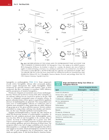
Fig. 43.5 MODIFICATIONS OF THE HEME AND ITS ENVIRONMENT THAT ACCOUNT FOR
TWO COMMON M HEMOGLOBINS. (A) Hemoglobin A has a His residue at the α58(E7) position.
(B) In hemoglobin M-Boston, the histidine is replaced by a tyrosine, the phenolic side chain of which is
capable of covalently binding to the heme iron, resulting in stabilization in the oxidized form. (C) HbA has
a Val residue at position β67(E11). (D) Hb M-Milwaukee has a glutamic acid substitution for the β67 valine.
The carboxylic side chain of the Glu forms a bond with iron, shifting the equilibrium toward the ferric state.
(Modified from Dickerson RE, Geis I: Hemoglobin: Structure, function, evolution, and pathology, Menlo Park, CA,
1983, Benjamin-Cummings. Copyright Irving Geis.)
hemoglobin to methemoglobin (Table 43.4). Some compounds TABLE Drugs and Chemicals Having Toxic Effects on
directly oxidize hemoglobin, whereas other compounds produce 43.4 Hemoglobin Molecule
reactive oxygen intermediates that oxidize hemoglobin. Nitrite
compounds are especially notorious and common. Some of these Observed Hemoglobin Derivative
compounds also have a propensity to exacerbate G6PD deficiency Agent Methemoglobin Sulfhemoglobin
and the precipitation of unstable hemoglobins.
Nitrates are a frequent environmental cause of toxic methemoglo- Acetanilid, phenacetin + +
binemia. Nitrates do not directly interact with either hemoglobin or Nitrites (ferric, amyl, sodium, + +
the reductase pathway but are converted to nitrites in the gut. Well potassium, nitroglycerin)
water is a frequently encountered source of excessive nitrates. In Trinitrotoluene, nitrobenzene + +
general, substantial intake of these agents is required before significant
amounts of methemoglobin are generated. Very young infants have Aniline, hydroxylamine + +
lower levels of methemoglobin reductase in erythrocytes and are dimethylamine
therefore more susceptible to these agents than are adults. However, Sulfanilamide + +
all age groups are at risk, given sufficient exposure. Systemic acidosis, p-Aminosalicylic acid +
particularly in young infants suffering from diarrhea and dehydra-
tion, can also cause clinically-significant methemoglobinemia. Dapsone +
Acquired methemoglobinemia is virtually the only situation in Primaquine, chloroquine +
which life-threatening amounts of methemoglobin accumulate. In Prilocaine, benzocaine, lidocaine +
general the only symptom produced when methemoglobin consti- +
tutes less than 30% of total hemoglobin is the cosmetic effect of Menadione, naphthoquinone
cyanosis. As levels of methemoglobin rise to greater than 30%, Naphthalene +
however, patients begin to exhibit symptoms of oxygen deprivation, Resorcinol +
such as malaise, giddiness, and other alterations of mental status. The Phenylhydrazine + +
symptoms reflect a true lack of oxygen availability at the tissue level.
Methemoglobin is a markedly left-shifted hemoglobin that delivers
little oxygen to the tissues. When methemoglobin accounts for more
than 50% of total hemoglobin, loss of consciousness, coma, and
death can rapidly ensue. At this level the blood is chocolate brown.