
Page 726 - Hematology_ Basic Principles and Practice ( PDFDrive )
P. 726
Chapter 43 Hemoglobin Variants Associated With Hemolytic Anemia, Altered Oxygen Affinity, and Methemoglobinemias 613
level is very high (>55% to 60%). The blood viscosity is then suffi- TABLE
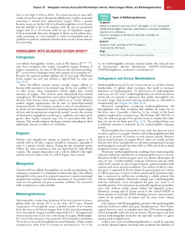
ciently elevated to require therapeutic phlebotomy. Carbon monoxide 43.3 Types of Methemoglobinemia
poisoning is treated with supplemental oxygen. When a patient
breathes room air, the half-life of carboxyhemoglobin is 4 to 6 hours, Congenital 3+ 2+
but the half-life is 40 to 80 minutes with the use of normobaric Defective enzymatic reduction of Fe -hemoglobin to Fe -hemoglobin
oxygen and 15 to 30 minutes with the use of hyperbaric oxygen. NADH-methemoglobin reductase (cytochrome-b 5 reductase) deficiency
Carbon monoxide detectors, designed to detect occult carbon mon- Cytochrome b 3 deficiency
oxide poisoning, are now required in many municipalities and are Abnormal hemoglobins resistant to enzymatic reduction (M
predicted to prevent numerous fatalities from occult carbon monox- hemoglobins)
ide poisoning. Acquired
2+
Excessive (toxic) oxidation of Fe -hemoglobin
Environmental chemicals
HEMOGLOBINS WITH DECREASED OXYGEN AFFINITY Drugs
NADH, Reduced form of nicotinamide adenine dinucleotide.
Pathogenesis
Low-affinity hemoglobin variants, such as Hb Kansas (β 102Asn→Thr ), by the methemoglobin reductase enzyme system (the reduced form
arise from mutations that impair hemoglobin-oxygen binding or of nicotinamide adenine dinucleotide [NADH]–dehydratase,
reduce cooperativity. In cases of Hb Kansas, the threonine position, [NADH]-diaphorase, erythrocyte cytochrome b 5 ).
102
94
β , cannot form a hydrogen bond with aspartic acid at position α .
Because this aspartate residue stabilizes the R (oxy) state, Hb Kansas
binds oxygen less well and exhibits a right-shifted P 50 value (see Pathogenesis and Clinical Manifestations
Fig. 43.4).
Most low-affinity variants possess enough oxygen affinity to Methemoglobinemias of clinical interest arise by one of three distinct
become fully saturated in the normal lung. At the low capillary PO 2 mechanisms: (1) globin chain mutations that result in increased
in other tissues, these hemoglobins deliver higher than normal formation of methemoglobin, (2) deficiencies of methemoglobin
amounts of oxygen. They become more desaturated than normal reductase, and (3) “toxic” methemoglobinemia, in which normal red
hemoglobins. Two abnormalities result from this high level of oxygen blood cells are exposed to substances that oxidize hemoglobin iron
delivery. First, because tissue oxygen delivery is so “overly” efficient, to such a degree that normal reducing mechanisms are subverted or
normal oxygen requirements can be met by lower-than-normal overwhelmed (see Chapter 44; Table 43.3).
hematocrit levels. This situation produces a state of “pseudoanemia,” Abnormal hemoglobins producing methemoglobinemia (M
in which the low hematocrit level is deceiving because both oxygen hemoglobins) arise from mutations that stabilize the heme iron in
delivery and the patients are completely normal. Second, the amount the ferric state. Classically a histidine in the vicinity of the heme
of desaturated hemoglobin circulating in capillaries and veins can be pocket is replaced by a tyrosine (e.g., Hb M-Iwate, β87 (F8) His →
greater than 5 g/dL. Cyanosis may thus be associated with these Tyr); the hydroxyl group of the tyrosine forms a complex that stabi-
variants. This usually ominous finding is entirely misleading in these lizes the iron in the ferric state (Fig. 43.5). The oxidized heme iron
individuals, because it reflects no morbidity. is relatively resistant to reduction by the methemoglobin reductase
system.
Methemoglobin has a brownish to blue color that does not revert
Diagnosis to red on exposure to oxygen. Patients with methemoglobinemia thus
appear to be cyanotic. In contrast to truly cyanotic people, however,
Patients with unexplained anemia or cyanosis who appear to be arterial partial pressure of oxygen (PaO 2 ) values are usually normal.
entirely well in all other respects should be evaluated, especially if Patients with these hemoglobins are otherwise asymptomatic because
there is a positive family history. Testing for the abnormal variant methemoglobin is usually less than 30% to 50%, the levels at which
follows the same reasoning as that just described for high-affinity symptoms become apparent.
variants. The oxygen dissociation curve will be shifted to the right, Hereditary methemoglobinemia resulting from methemoglobin
and the numeric value of the P 50 will be higher than normal. reductase deficiency (cytochrome-b 5 reductase deficiency) is very rare.
Mutations in the b 5 reductase gene cause two distinct phenotypes. In
cases of type I methemoglobin reductase deficiency, patients suffer
Management solely from cyanosis; in cases of type II disease, patients manifest both
cyanosis and severe mental retardation. One isoform of the b 5 reduc-
Patients with low-affinity hemoglobins are usually asymptomatic. No tase gene is expressed in diverse tissues for participation in a variety
treatment is required. It is important to document that a low-affinity of cellular processes. A second isoform, produced by alternative splic-
hemoglobin is the cause of an apparent anemia or cyanosis to preempt ing, is expressed in erythrocytes, producing a soluble protein that
inappropriate workups and provide reassurance to the patient. Cya- reduces methemoglobin. Mutations causing type I methemoglobin
nosis in some patients can pose a cosmetic problem, but correction reductase deficiency occur throughout the gene and result in an
with transfusions is rarely justified. unstable protein. Such mutations are primarily significant in erythro-
cytes that, without nuclei, cannot replace the degraded protein.
Mutations causing type II disease occur in the critical NADH or
Methemoglobinemias flavin adenine dinucleotide (FAD)–binding domains, causing inacti-
vation of the protein in all tissues and the more severe clinical
Methemoglobin results from oxidation of the iron moieties in hemo- phenotype.
3+
2+
globin from the ferrous (Fe ) to the ferric (Fe ) state. Normal Like patients with M hemoglobins, patients with methemoglobin
oxygenation of hemoglobin causes a partial transfer of an electron reductase deficiency exhibit slate-gray “pseudocyanosis.” Even homo-
from the iron to the bound oxygen. Iron in this state thus resembles zygotes, however, rarely accumulate more than 25% methemoglobin,
−
ferric iron and the oxygen resembles superoxide (O 2 ). Deoxygenation a level compatible with minimal symptoms. Heterozygotes can have
returns the electron to the iron, with release of oxygen. Methemoglo- normal methemoglobin levels but are especially sensitive to agents
bin forms if the electron is not returned. Methemoglobin constitutes causing methemoglobinemia.
3% or less of the total hemoglobin in normal humans. Under normal A third toxic form of methemoglobinemia is caused by exposure
circumstances, these levels in humans are maintained at 1% or less to certain chemical agents and drugs that accelerate the oxidation of