
Page 742 - Hematology_ Basic Principles and Practice ( PDFDrive )
P. 742
Chapter 45 Red Blood Cell Membrane Disorders 629
Spectrin/ankyrin Hemolysis
deficiency ↓ pH
↑ Macrophage
contact
Release of microvesicles
Splenic Splenic Further loss
trapping, conditioning of membrane
erythrostasis
Band 3/protein 4.2
deficiency
Spherocytes
Release of microvesicles
Microspherocytes
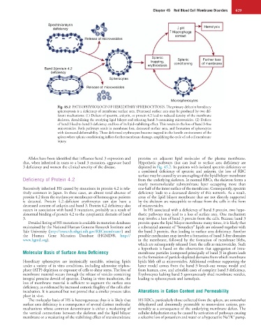
Fig. 45.2 PATHOPHYSIOLOGY OF HEREDITARY SPHEROCYTOSIS. The primary defect in hereditary
spherocytosis is a deficiency of membrane surface area. Decreased surface area may be produced by two dif-
ferent mechanisms: (1) Defects of spectrin, ankyrin, or protein 4.2 lead to reduced density of the membrane
skeleton, destabilizing the overlying lipid bilayer and releasing band 3-containing microvesicles. (2) Defects
of band 3 lead to band 3 deficiency and loss of its lipid-stabilizing effect. This results in the loss of band 3-free
microvesicles. Both pathways result in membrane loss, decreased surface area, and formation of spherocytes
with decreased deformability. These deformed erythrocytes become trapped in the hostile environment of the
spleen where splenic conditioning inflicts further membrane damage, amplifying the cycle of red cell membrane
injury.
Alleles have been identified that influence band 3 expression and proteins on adjacent lipid molecules of the plasma membrane.
that, when inherited in trans to a band 3 mutation, aggravate band Hypothetic pathways that can lead to surface area deficiency are
3 deficiency and worsen the clinical severity of the disease. depicted in Fig. 45.2. In patients with isolated spectrin deficiency or
a combined deficiency of spectrin and ankyrin, the loss of RBC
surface may be caused by an uncoupling of the lipid bilayer membrane
Deficiency of Protein 4.2 from the underlying skeleton. In normal RBCs, the skeleton forms a
nearly monomolecular submembrane layer occupying more than
Recessively inherited HS caused by mutations in protein 4.2 is rela- one-half of the inner surface of the membrane. Consequently, spectrin
tively common in Japan. In these cases, an almost total absence of deficiency leads to a decreased density of this network. As a result,
protein 4.2 from the erythrocyte membranes of homozygous patients areas of the lipid bilayer membrane that are not directly supported
is detected. Protein 4.2–deficient erythrocytes can also have a by the skeleton are susceptible to release from the cells in the form
decreased content of ankyrin and band 3. Protein 4.2 deficiency also of microvesicles.
occurs in association with band 3 mutations, probably as a result of In HS associated with a deficiency of band 3 protein, two hypo-
abnormal binding of protein 4.2 to the cytoplasmic domain of band thetic pathways may lead to a loss of surface area. One mechanism
3. may involve a loss of band 3 protein from the cells. Because band 3
Detailed listing of HS mutations is available in mutation databases protein spans the lipid bilayer membrane many times, it is likely that
maintained by the National Human Genome Research Institute and a substantial amount of “boundary” lipids are released together with
Yale University (http://research.nhgri.nih.gov/RBCmembrane/) and the band 3 protein, thus leading to surface area deficiency. Another
the Human Gene Mutation Database (HGMD®, http:// possible mechanism may involve a formation of band 3-free domains
www.hgmd.org). in the membrane, followed by the formation of membrane blebs,
which are subsequently released from the cells as microvesicles. Such
a hypothesis is based on the observation that aggregation of intra-
Molecular Basis of Surface Area Deficiency membrane particles (composed principally of band 3) in ghosts leads
to the formation of particle-depleted domains from which membrane
Hereditary spherocytes are intrinsically unstable, releasing lipids lipids bleb off as microvesicles. Additional evidence supporting the
under a variety of in vitro conditions, including adenosine triphos- latter model comes from the band 3 knock-out mouse model and
phate (ATP) depletion or exposure of cells to shear stress. The loss of from human, cow, and zebrafish cases of complete band 3 deficiency.
membrane material occurs through the release of vesicles containing Erythrocytes lacking band 3 spontaneously shed membrane vesicles,
integral proteins devoid of spectrin. During in vitro incubation, the leading to spherocytosis and hemolysis.
loss of membrane material is sufficient to augment the surface area
deficiency, as evidenced by increased osmotic fragility of the cells after
incubation. It is assumed but not proved that a similar process takes Alterations in Cation Content and Permeability
place in vivo.
The molecular basis of HS is heterogeneous; thus it is likely that HS RBCs, particularly those collected from the spleen, are somewhat
surface area deficiency is a consequence of several distinct molecular dehydrated and abnormally permeable to monovalent cations, pre-
mechanisms whose common denominator is either a weakening of sumably as a consequence of the underlying membrane defect. The
the vertical connections between the skeleton and the lipid bilayer cellular dehydration may be caused by activation of pathways causing
+
+
membrane or a weakening of the stabilizing effect of transmembrane a selective loss of potassium and water or a hyperactive Na /K pump.