
Page 740 - Hematology_ Basic Principles and Practice ( PDFDrive )
P. 740
Chapter 45 Red Blood Cell Membrane Disorders 627
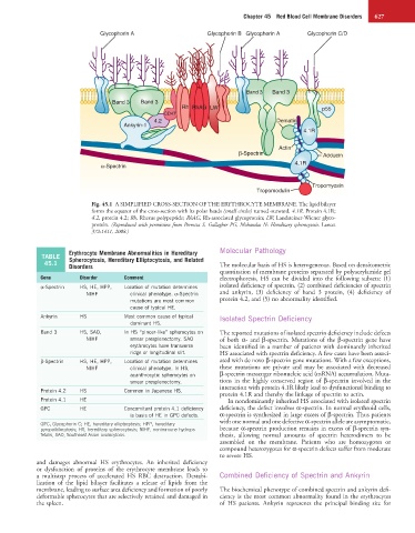
Glycophorin A Glycophorin B Glycophorin A Glycophorin C/D
Band 3 Band 3
Band 3 Band 3
Rh RhAG LW p55
CD47
4.2 Dematin
Ankyrin-1
4.1R
Actin
β-Spectrin Adducin
4.1R
α-Spectrin
Tropomyosin
Tropomodulin
Fig. 45.1 A SIMPLIFIED CROSS-SECTION OF THE ERYTHROCYTE MEMBRANE. The lipid bilayer
forms the equator of the cross-section with its polar heads (small circles) turned outward. 4.1R, Protein 4.1R;
4.2, protein 4.2; Rh, Rhesus polypeptide; RhAG, Rh-associated glycoprotein; LW, Landsteiner-Wiener glyco-
protein. (Reproduced with permission from Perrotta S, Gallagher PG, Mohandas N: Hereditary spherocytosis. Lancet
372:1411, 2008.)
Erythrocyte Membrane Abnormalities in Hereditary Molecular Pathology
TABLE Spherocytosis, Hereditary Elliptocytosis, and Related
45.1
Disorders The molecular basis of HS is heterogeneous. Based on densitometric
quantitation of membrane proteins separated by polyacrylamide gel
Gene Disorder Comment electrophoresis, HS can be divided into the following subsets: (1)
α-Spectrin HS, HE, HPP, Location of mutation determines isolated deficiency of spectrin, (2) combined deficiencies of spectrin
NIHF clinical phenotype. α-Spectrin and ankyrin, (3) deficiency of band 3 protein, (4) deficiency of
mutations are most common protein 4.2, and (5) no abnormality identified.
cause of typical HE.
Ankyrin HS Most common cause of typical Isolated Spectrin Deficiency
dominant HS.
Band 3 HS, SAO, In HS “pincer-like” spherocytes on The reported mutations of isolated spectrin deficiency include defects
NIHF smear presplenectomy. SAO of both α- and β-spectrin. Mutations of the β-spectrin gene have
erythrocytes have transverse been identified in a number of patients with dominantly inherited
ridge or longitudinal slit. HS associated with spectrin deficiency. A few cases have been associ-
β-Spectrin HS, HE, HPP, Location of mutation determines ated with de novo β-spectrin gene mutations. With a few exceptions,
NIHF clinical phenotype. In HS, these mutations are private and may be associated with decreased
acanthrocytic spherocytes on β-spectrin messenger ribonucleic acid (mRNA) accumulation. Muta-
smear presplenectomy. tions in the highly conserved region of β-spectrin involved in the
interaction with protein 4.1R likely lead to dysfunctional binding to
Protein 4.2 HS Common in Japanese HS. protein 4.1R and thereby the linkage of spectrin to actin.
Protein 4.1 HE In nondominantly inherited HS associated with isolated spectrin
GPC HE Concomitant protein 4.1 deficiency deficiency, the defect involves α-spectrin. In normal erythroid cells,
is basis of HE in GPC defects. α-spectrin is synthesized in large excess of β-spectrin. Thus patients
with one normal and one defective α-spectrin allele are asymptomatic,
GPC, Glycophorin C; HE, hereditary elliptocytosis; HPP, hereditary
pyropoikilocytosis; HS, hereditary spherocytosis; NIHF, nonimmune hydrops because α-spectrin production remains in excess of β-spectrin syn-
fetalis, SAO, Southeast Asian ovalocytosis. thesis, allowing normal amounts of spectrin heterodimers to be
assembled on the membrane. Patients who are homozygotes or
compound heterozygotes for α-spectrin defects suffer from moderate
to severe HS.
and damages abnormal HS erythrocytes. An inherited deficiency
or dysfunction of proteins of the erythrocyte membrane leads to
a multistep process of accelerated HS RBC destruction. Destabi- Combined Deficiency of Spectrin and Ankyrin
lization of the lipid bilayer facilitates a release of lipids from the
membrane, leading to surface area deficiency and formation of poorly The biochemical phenotype of combined spectrin and ankyrin defi-
deformable spherocytes that are selectively retained and damaged in ciency is the most common abnormality found in the erythrocytes
the spleen. of HS patients. Ankyrin represents the principal binding site for