
Page 744 - Hematology_ Basic Principles and Practice ( PDFDrive )
P. 744
Chapter 45 Red Blood Cell Membrane Disorders 631
Laboratory Manifestations smear evaluation; the cells appear as “fat” disks rather than as true
spherocytes.
Most HS patients have mild to moderate anemia or no anemia at all, Additional morphologic features have been described in some HS
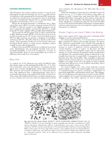
reflecting the fact that the hemolytic rate can be very mild and that patients (see Fig. 45.3). A subset of HS patients whose RBCs are
the hemolysis is fully compensated for by increased RBC production, deficient in band 3 protein have some pincer-like RBCs on the
as evidenced by reticulocytosis. Some patients, however, particularly peripheral blood film, a finding that is both sensitive and specific for
those with nondominantly inherited HS, are severely anemic, with this HS subset. These pincer-like cells disappear after splenectomy.
hemoglobin concentrations as low as 4 to 6 g/dL. Surface spiculations or acanthocytic spherocytes have been described
Despite the increased percentage of reticulocytes with a larger in cases of HS associated with defects in β-spectrin. Frequent sphero-
volume than mature RBCs, the mean corpuscular volume (MCV) of ovalocytes and stomatocytes have been reported in Japanese patients
HS RBCs is often low normal or even slightly decreased, and the with protein 4.2 deficiency.
mean corpuscular hemoglobin concentration (MCHC) is usually
increased (>35 g/dL), together reflecting mild cellular dehydration.
The finding of an MCHC greater than 35.4 g/dL combined with Osmotic Fragility and Eosin-5′-Maleimide Binding
an RBC distribution width (RDW) <14% has been found to be an
excellent screening test for HS. Another screening method measures Both osmotic fragility (OF) testing and eosin-5′-maleimide (EMA)
MCV by light scattering and provides a histogram of hyperdense binding are used in evaluating HS patients.
erythrocytes (MCHC >40 g/dL) claimed to identify nearly all HS The osmotic fragility test (Fig. 45.4) measures the in vitro lysis of
patients. These hyperdense erythrocytes can be detected with newer RBCs suspended in solutions of decreasing osmolarity. The normal
laser-based blood counters or using aperture impedance analysis RBC membrane is unstretchable and is virtually freely permeable to
available in many clinical laboratories. water. Thus the cell behaves as a nearly perfect osmometer in that it
Evidence of accelerated RBC destruction, as indicated by increased increases its volume in hypotonic solutions progressively until a
lactate dehydrogenase and unconjugated bilirubin levels and by “critical hemolytic volume” is reached. At this point, the RBC
decreased haptoglobin, as well as by reticulocytosis, is present in membrane ruptures, and hemoglobin escapes into the supernatant
typical HS patients. However, these abnormalities can be absent in solution. As a result of the loss of membrane and the ensuing surface
individuals with a mild form of the disease. area deficiency, the critical hemolytic volume of spherocytes is con-
siderably lower than that of normal RBCs. Consequently, these cells
hemolyze more than normal RBCs when suspended in hypotonic
Blood Film sodium chloride solutions. However, a finding of increased osmotic
fragility is not unique to HS and is also present in other conditions
In a typical case of HS, spherocytes are readily identified by their associated with spherocytosis on the peripheral blood film, such as
characteristic shape on the peripheral blood film (Fig. 45.3). They autoimmune hemolytic anemia.
lack central pallor, their mean cell diameter is decreased, and they The osmotic fragility curve often reveals uniformly increased
appear more intensely hemoglobinized, which reflects both altered osmotic fragility. A “tail” of the osmotic fragility curve can be present
RBC geometry and increased cell density. In a three-dimensional in nonsplenectomized HS patients, indicating a subpopulation of
view, some spherocytes have a spherostomatocytic shape that is particularly fragile RBCs conditioned by splenic stasis. This subpopu-
occasionally appreciated on the peripheral blood film. In mild forms lation of cells disappears after splenectomy. In patients with mild HS,
of the disease, the peripheral blood smear can appear normal because osmotic fragility can be normal and abnormalities can be found only
the loss of surface area can be too small to be appreciated by blood after incubation that further augments the loss of surface area;
A B
C D
Fig. 45.3 BLOOD FILMS FROM PATIENTS WITH HEREDITARY SPHEROCYTOSIS (HS) OF
VARYING SEVERITY. (A) Two blood films of typical moderately severe HS with a mild deficiency of RBC
spectrin and ankyrin. Although many cells have a spheroidal shape, some retain a central concavity. (B) HS
with pincer-like RBCs (arrows), as typically seen in HS associated with band 3 deficiency. Occasional spiculated
RBCs are also present. (C) Severe atypical HS caused by a severe combined spectrin and ankyrin deficiency.
In addition to spherocytes, many cells have irregular contour. (D) HS with isolated spectrin deficiency caused
by a β-spectrin mutation. Some of the spherocytes have prominent surface projections resembling
sphero-acanthocytes.