
Page 803 - Hematology_ Basic Principles and Practice ( PDFDrive )
P. 803
Chapter 49 Lymphocytosis, Lymphocytopenia, Hypergammaglobulinemia, and Hypogammaglobulinemia 689
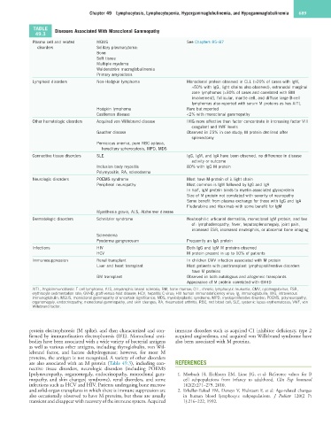
TABLE Diseases Associated With Monoclonal Gammopathy
49.3
Plasma cell and related MGUS See Chapters 85–87
disorders Solitary plasmacytoma:
Bone
Soft tissue
Multiple myeloma
Waldenström macroglobulinemia
Primary amyloidosis
Lymphoid disorders Non-Hodgkin lymphoma Monoclonal protein observed in CLL (>20% of cases with IgM,
≈50% with IgG, light chains also observed), extranodal marginal
zone lymphomas (>30% of cases and correlated with BM
involvement), follicular, mantle cell, and diffuse large B-cell
lymphomas also reported with serum M proteins as has AITL
Hodgkin lymphoma Rare but reported
Castleman disease <2% with monoclonal gammopathy
Other hematologic disorders Acquired von Willebrand disease IVIG more effective than factor concentrate in increasing factor VIII
coagulant and VWF levels
Gaucher disease Observed in 25% in one study; M protein declined after
splenectomy
Pernicious anemia, pure RBC aplasia,
hereditary spherocytosis, MPD, MDS
Connective tissue disorders SLE IgG, IgM, and IgA have been observed, no difference in disease
activity or outcome
Inclusion body myositis 80% with IgG M protein
Polymyositis, RA, scleroderma
Neurologic disorders POEMS syndrome Most have M-protein of λ light chain
Peripheral neuropathy Most common is IgM followed by IgG and IgA
In half, IgM protein binds to myelin-associated glycoprotein
Size of M protein not correlated with severity of neuropathy
Some benefit from plasma exchange for those with IgG and IgA
Fludarabine and rituximab with some benefit for IgM
Myasthenia gravis, ALS, Alzheimer disease
Dermatologic disorders Schnitzler syndrome Neutrophilic urticarial dermatitis, monoclonal IgM protein, and two
of: lymphadenopathy, fever, hepatosplenomegaly, joint pain,
increased ESR, increased neutrophils, or abnormal bone imaging
Scleredema
Pyoderma gangrenosum Frequently an IgA protein
Infections HIV Both IgG and IgM M proteins observed
HCV M protein present in up to 10% of patients
Immunosuppression Renal transplant In children CMV infection associated with M protein
Liver and heart transplant Most patients with posttransplant lymphoproliferative disorders
have M proteins
BM transplant Observed in both autologous and allogeneic transplants
Appearance of M protein correlated with GVHD
AITL, Angioimmunoblastic T-cell lymphoma; ALS, amyotrophic lateral sclerosis; BM, bone marrow; CLL, chronic lymphocytic leukemia; CMV, cytomegalovirus; ESR,
erythrocyte sedimentation rate; GVHD, graft-versus-host disease; HCV, hepatitis C virus; HIV human immunodeficiency virus; Ig, immunoglobulin; IVIG, intravenous
immunoglobulin; MGUS, monoclonal gammopathy of uncertain significance; MDS, myelodysplastic syndrome; MPD, myeloproliferative disorder; POEMS, polyneuropathy,
organomegaly, endocrinopathy, monoclonal gammopathy, and skin changes; RA, rheumatoid arthritis; RBC, red blood cell; SLE, systemic lupus erythematosus; VWF, von
Willebrand factor.
protein electrophoresis (M spike), and then characterized and con- immune disorders such as acquired C1 inhibitor deficiency, type 2
firmed by immunofixation electrophoresis (IFE). Monoclonal anti- acquired angioedema, and acquired von Willebrand syndrome have
bodies have been associated with a wide variety of bacterial antigens also been associated with M proteins.
as well as various other antigens, including thyroglobulin, von Wil-
lebrand factor, and lactate dehydrogenase; however, for most M
proteins, the antigen is not recognized. A variety of other disorders
are also associated with an M protein (Table 49.3), including con- REFERENCES
nective tissue disorders, neurologic disorders (including POEMS
[polyneuropathy, organomegaly, endocrinopathy, monoclonal gam- 1. Morbach H, Eichhorn EM, Liese JG, et al: Reference values for B
mopathy, and skin changes] syndrome), renal disorders, and some cell subpopulations from infancy to adulthood. Clin Exp Immunol
infections such as HCV and HIV. Patients undergoing bone marrow 162(2):271–279, 2010.
and solid-organ transplants in which there is immune suppression are 2. Erkeller-Yuksel FM, Deneys V, Hulstaert F, et al: Age-related changes
also occasionally observed to have M proteins, but these are usually in human blood lymphocyte subpopulations. J Pediatr 120(2 Pt
transient and disappear with recovery of the immune system. Acquired 1):216–222, 1992.