
Page 1048 - Williams Hematology ( PDFDrive )
P. 1048
1022 Part VII: Neutrophils, Eosinophils, Basophils, and Mast Cells Chapter 66: Disorders of Neutrophil Function 1023
such as S. aureus, Pseudomonas spp. and other Gram-negative enteric responses, for example, to repeat vaccination with tetanus toxoid, diph-
rods, or Candida spp. Patients with the moderate phenotype have fewer theria toxoid, and polio virus.
and less-severe infections. Infectious susceptibility and impaired wound Patients with LAD-1 usually have blood neutrophil counts of 15 to
healing are related to diminished or delayed infiltration of neutrophils 60 × 10 /L. However, during infectious episodes, they commonly have
9
and monocytes into extravascular inflammatory sites. In all patients neutrophil counts in excess of 100 × 10 /L and sometimes as high as 160 ×
9
9
surviving infancy, severe progressive generalized periodontitis is pres- 10 /L. Granulocytic hyperplasia is a feature of the marrow examina-
ent. Individuals who are clinically well, but who are heterozygous car- tion which may relate to excessive production of IL-17 and G-CSF as
riers of LAD have been identified. Their stimulated neutrophils express a result of decreased uptake of apoptotic neutrophils by tissue macro-
approximately 50 percent of the normal amount of the Mac-1 α sub- phages. 319,330 Despite elevated blood counts, there is a paucity of neu-
309
unit and the common β subunit. The diagnosis of LAD-1 should be trophils in inflammatory skin windows and biopsies of infected tissues.
considered in infants with a paucity of neutrophils at sites of infection Differential Diagnosis Eight patients (four Arab, two Turkish,
despite blood neutrophilia and have a history of delayed separation of one Pakistani, one Brazilian) who had neutrophilia, recurrent bacterial
the umbilical cord. infections, and an inability to form pus have been described. 13,331,332 The
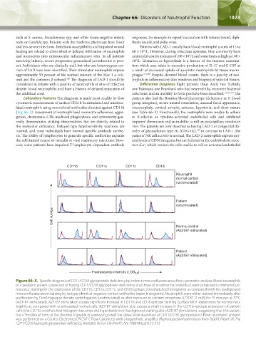
Laboratory Features The diagnosis is made most readily by flow patients also had the Bombay blood phenotype (deficiency in H blood
cytometric measurement of surface CD11b in stimulated and unstimu- group integrins), severe mental retardation, unusual facial appearance,
lated neutrophils using monoclonal antibodies directed against CD11b microcephaly, cortical atrophy, seizures, hypotonia, and short stature
(Fig. 66–5). Assessment of neutrophil and monocyte adherence, aggre- (see Table 66–2). Functionally, the neutrophils were unable to adhere
gation, chemotaxis, C3bi-mediated phagocytosis, and cytotoxicity gen- to E-selectin or cytokine-activated endothelial cells and exhibited
erally demonstrates striking abnormalities that are directly related to impaired chemotaxis and an inability to roll on postcapillary venules in
the molecular deficiency. Delayed-type hypersensitivity reactions are vivo. The patients are now classified as having LAD-2 or congenital dis-
333
normal, and most individuals have normal specific antibody synthe- order of glycosylation type IIc (CDG-IIc). In contrast to LAD-1, the
sis. The ability of lymphocytes to generate specific antibodies explains patients’ NK cell activity is normal. The LAD-2 neutrophils express nor-
the self-limited course of varicella or viral respiratory infections. How- mal levels of CD18 integrins, but are deficient in the carbohydrate struc-
ever, some patients have impaired T-lymphocyte–dependent antibody ture sLe , which renders the cells unable to roll on activated endothelial
x
CD11b CD11a CD11c CD18
Neutrophil
normal control
(unstimulated)
Patient
(unstimulated)
Cell number
Normal control
(A23187-stimulated)
Patient
(A23187-stimulated)
Fluorescence intensity (LOG )
10
Figure 66–5. Specific diagnosis of CD11/CD18 glycoprotein deficiency by indirect immunofluorescence flow cytometric analysis. Blood neutrophils
of a pediatric patient suspected of having CD11/CD18 glycoprotein deficiency and those of an abnormal individual were subjected to immunofluo-
rescence staining for the expression of the CD11b, CD11a, CD11c, and CD18 epitope (crosshatched histograms) as compared with the background
immunofluorescence staining by isotype-identical negative-control antibodies (open histograms). Neutrophils were either stained immediately after
purification by Ficoll-Hypaque density centrifugation (unstimulated) or after exposure to calcium ionophore A23187 (1 mM) for 15 minutes at 37°C
(A23187-stimulated). A23187 stimulation causes significant increase in CD11b and CD18 epitope staining (surface MO1 expression) by normal neu-
trophils as compared with unstimulated normal cells. A23187 stimulation also causes a small increase in the CD11b-epitope expression of patient
cells (the CD11b crosshatched histogram becomes distinguishable from background staining after A23187 stimulation), suggesting that this patient
has a “moderate” form of the disorder (capable of expressing small but detectable quantities of CD11/CD18 glycoproteins). Flow cytometric analysis
was performed on a Coulter Electronics EPICS F C Flow Cytometer with a logarithmic amplifier. (Reproduced with permission from Todd R, Freyer DR: The
CD11/CD18 leukocyte glycoprotein deficiency, Hematol Oncol Clin North Am 1988 Mar;2(1):13-31.)
Kaushansky_chapter 66_p1005-1042.indd 1023 9/21/15 10:48 AM