
Page 110 - Williams Hematology ( PDFDrive )
P. 110
86 Part II: The Organization of the Lymphohematopoietic Tissues Chapter 6: The Organization and Structure of Lymphoid Tissues 87
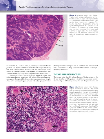
Figure 6–1. Normal human infant thymus.
The thymus is surrounded by dense connec-
tive tissue capsule (Cap). It is organized into
adjacent lobules separated by capsular con-
nective tissue extensions or trabeculae. The
lobules each have a dense cortex (C) and a
lighter staining medulla (M). The medulla is a
continuous tissue surrounded by the cortex
that extends throughout the thymus, and
it cannot be appreciated in a single section.
(Reproduced with permission from Lichtman’s
Atlas of Hematology, www.accessmedicine.
com.)
M
C
Cap
as interleukin (IL)-7. In addition, at primarily the corticomedullary thymocytes. This also may be seen in conditions that are associated
5
11
junction, the thymus contains marrow-derived antigen-presenting with increases in circulating glucocorticoid hormones, for example,
cells, mostly interdigitating dendritic cells and macrophages. Scat- pregnancy or stress. 14,15
tered B cells are also present in the thymus, and these interact with
maturing thymocytes and potentially regulate T-cell development. 12,13
After puberty, thymic involution begins within the cortex. This THYMIC IMMUNE FUNCTION
region may disappear completely with aging, while medullary rem- The thymus is the site of T-cell development. The importance of the
nants persist throughout life. Glucocorticoids also may induce atrophy thymus is underscored by patients with DiGeorge syndrome, or chro-
of the cortex secondary to glucocorticoid-induced apoptosis of cortical mosome 22q11.2 deletion syndrome, who lack the genes required for
Figure 6–2. Normal human infant thymus.
Higher magnification. Medulla. The arrows
indicate thymic corpuscles (synonymous with
Hassall corpuscles). They are composed of
tightly packed, concentrically arranged, type
IV endothelioreticular cells with flattened
nuclei. The central mass is composed of ker-
atinized cells. In addition to thymic corpus-
cles and the mass of small densely stained T
lymphocytes, the medulla contains scattered,
larger, type V epithelioreticular cells with their
light nuclei, dark nucleolus, and eosinophilic
cytoplasm, evident on this section. (Repro-
duced with permission from Lichtman’s Atlas of
Hematology, www.accessmedicine.com.)
Kaushansky_chapter 06_p0085-0096.indd 86 17/09/15 5:52 pm