
Page 111 - Williams Hematology ( PDFDrive )
P. 111
86 Part II: The Organization of the Lymphohematopoietic Tissues Chapter 6: The Organization and Structure of Lymphoid Tissues 87
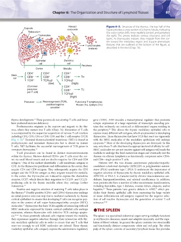
Hassall Figure 6–3. Structure of the thymus. The top half of the
Capsule
corpuscle figure provides a cross section of a thymic lobule, indicating
Trabeculum the outer cortex (left), inner medulla (center), and periphery
Thymocytes (far right). The arrows indicate various structures and cell
types. As thymocytes mature, they migrate from the cor-
tex toward the medullary region and acquire phenotypic
T lymphocytes features that are outlined at the bottom of the figure, as
(mature) described in the text (Chap. 74).
Epithelial Dendritic
cell cell
Periphery
Cortex Medulla
cell { CD4 – CD4 + CD4 +
Stem CD8 – { CD8 + { CD8 – TH
TCR – TCRαβ TCR αβ
(low) (high)
(Common CD4 –
precursor { CD8 + TC
pre-T cell)
TCR αβ
(high)
Precursor Rearrangement (TCR), Functional T lymphocytes
positive/negative Helper (Th), cytolytic (TC)
selection
thymic development. These patients do not develop T cells and hence gene (AIRE). AIRE encodes a transcriptional regulator that promotes
16
have profound immune deficiency. ectopic expression of a large repertoire of transcripts encoding pro-
Prothymocytes originate in the marrow and migrate to the thy- teins that ordinarily are restricted to differentiated organs residing in
mus, where they mature into T cells (Chap. 76). Maturation of T cells the periphery. This allows the thymic medullary epithelial cells to
25
is accompanied by the sequential acquisition of various T-cell markers express many different self-antigens, which are presented to developing
including CD2, CD3, CD4 or CD8, CD5, and the T-cell receptor (TCR) thymocytes. Those thymocytes that have TCR that react too vigorously
(Fig. 6–3). Terminal deoxynucleotidyl transferase (TdT) is found in with the MHC molecules of the medullary epithelium will undergo
17
prothymocytes and immature thymocytes but is absent in mature apoptosis. Most of the developing thymocytes are destroyed. In this
23
T cells. TdT facilitates the successful rearrangement of TCR genes in way, only those T cells that have the appropriate level of affinity for self-
immature thymocytes. 18 MHC molecules yet are not reactive against self antigens will reach the
T-cell precursors can be found in distinct microenvironments medulla to undergo the final maturation stages and eventually exit the
within the thymus. Marrow-derived CD34+ pre-T cells enter the cor- thymus via efferent lymphatics as functionally competent naïve CD4+
tex via small blood vessels and are double-negative for CD4 and CD8 and CD8+ single-positive T cells.
antigens. One of the earliest identifiable T-cell membrane antigens is Patients with the rare disease autoimmune polyendocrinopathy-
1
CD2. As the thymocytes proliferate and differentiate in the cortex, they candidiasis-ectodermal dystrophy (APECED) or polyglandular autoim-
acquire CD4 and CD8 antigens. They subsequently acquire the CD3 mune (PGA) syndrome type I (PGA I) underscore the importance of
antigen and the TCR for antigen as they migrate toward the medulla. negative selection of thymocytes by thymic medullary epithelial cells.
In the cortex, the thymocytes are induced to express the chemokine APECED, or PGA I, is characterized by chronic mucocutaneous can-
receptor, CCR7, which directs their migration to CCL19- and CCL21- didiasis, hypoparathyroidism, and adrenal insufficiency. In addition,
producing cells in the thymic medulla where they undergo further most patients also have a number of other autoimmune manifestations,
maturation. 19 including thyroiditis, type 1 diabetes, ovarian failure, alopecia, and/or
Positive and negative selection of maturing T cells takes place in hepatitis. These patients have genetic defects in AIRE, which pre-
27
26
the thymus. Double-positive (CD4+ and CD8+) thymocytes undergo cludes their thymic epithelial cells from expressing the large variety
20
an initial positive selection step that is mediated exclusively by thymic of tissue differentiation self-antigens required for the negative selec-
cortical epithelium to ensure that developing T cells can recognize pep- tion of self-reactive thymocytes and the generation of central T-cell
tides in the context of self major histocompatibility complex (MHC) tolerance. 25,28
molecules. Thymocytes that have TCRs capable of interacting with self
21
MHC molecules expressed by thymic cortical epithelial cells undergo THE SPLEEN
expansion, whereas thymocytes with defective TCR undergo apopto-
sis. 22–24 As these positively selected cells migrate toward the medulla, The spleen is a specialized abdominal organ serving multiple functions
they experience negative selection through their interaction with thy- in erythrocyte clearance, innate and adaptive immunity, and the regu-
mic medullary epithelial cells in order to ensure that any T cells that lation of blood volume. In general the spleen contains two structurally
react too strongly to self MHC molecules are deleted. These thymic and functionally distinct components: white and red pulp. The white
medullary epithelial cells uniquely express the autoimmune regulatory pulp of the spleen consists of secondary lymphoid tissue that provides
Kaushansky_chapter 06_p0085-0096.indd 87 17/09/15 5:53 pm