
Page 1163 - Williams Hematology ( PDFDrive )
P. 1163
1138 Part IX: Lymphocytes and Plasma Cells
A B C D
E F G H
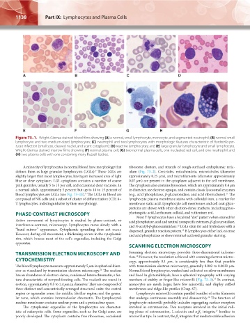
Figure 73–1. Wright-Giemsa stained blood films showing (A) a normal, small lymphocyte, monocyte, and segmented neutrophil; (B) normal small
lymphocyte and two medium-sized lymphocytes; (C) neutrophil and two lymphocytes with morphologic features characteristic of Bordetella per-
tussis infection (small size, cleaved nuclei, and scant cytoplasm); (D) reactive lymphocytes; and (E) large granular lymphocyte and small lymphocyte.
Wright-Giemsa stained marrow films showing (F) normal plasma cell; (G) two normal plasma cells, one nucleated red cell, and one neutrophil; and
(H) two plasma cells with one containing many Russell bodies.
A minority of lymphocytes in normal blood have morphology that ribosome clusters, and strands of rough-surfaced endoplasmic retic-
defines them as large granular lymphocytes (LGLs). These LGLs are ulum (Fig. 73–2). Centrioles, mitochondria, microtubules (diameter
9
slightly larger than most lymphocytes, having an increased area of light approximately 0.25 μm), and microfilaments (diameter approximately
blue or clear cytoplasm. LGL cytoplasm contains a number of coarse 0.07 μm) are present in the cytoplasm adjacent to the cell membrane.
pink granules, usually 5 to 15 per cell, and occasional clear vacuoles. In The cytoplasm also contains lysosomes, which are approximately 0.4 μm
a normal adult, approximately 5 percent but up to 10 to 15 percent of in diameter, are electron opaque, and contain classic lysosomal enzymes
blood lymphocytes are LGLs (see Fig. 73–1E). The LGLs in blood are (e.g., acid phosphatase, β-glucuronidase, and acid ribonuclease). The
11
9
composed of NK cells and a subset of cluster of differentiation (CD) 8+ lymphocyte plasma membrane stains with colloidal iron, a marker for
T lymphocytes, indistinguishable by their morphology. membrane sialic acid. Lymphocyte cell membranes and cell coat glyco-
proteins are shown with other electron-dense markers, including phos-
PHASE-CONTRAST MICROSCOPY photungstic acid, lanthanum colloid, and ruthenium red.
Most T lymphocytes have a localized “dot” pattern when stained for
Active movement of lymphocytes is studied by phase-contrast, or acid phosphatase, acid and neutral nonspecific esterases, β-glucuronidase,
interference-contrast, microscopy. Lymphocytes move slowly with a and N-acetyl-β-glucosaminidase. LGLs stain for acid hydrolases with a
12
“hand mirror” appearance. Cytoplasmic spreading does not occur. dispersed, granular reaction pattern. B lymphocytes either lack esterase
13
However, during cell movement, a thickening occurs in the cytoplasmic and acid phosphatase or show minimal scattered granular staining.
rim, which houses most of the cell’s organelles, including the Golgi
apparatus.
SCANNING ELECTRON MICROSCOPY
TRANSMISSION ELECTRON MICROSCOPY AND Scanning electron microscopy provides three-dimensional informa-
14
CYTOCHEMISTRY tion. However, the resolution achieved with scanning electron micros-
copy, approximately 0.1 μm, is considerably less than that possible
The blood lymphocyte measures approximately 5 μm in spherical diam- with transmission electron microscopy, generally 0.002 to 0.0039 μm.
10
eter as visualized by transmission electron microscopy. The nucleus Normal blood lymphocytes, washed and collected on silver membranes
has an abundance of electron-dense, condensed heterochromatin, a fea- and fixed in glutaraldehyde, have a spherical topography with varying
15
ture characteristic of nonproliferating cells. The nucleoli are round in numbers of stubby or finger-like microvilli (Fig. 73–3). In contrast,
section, approximately 0.5 to 1.4 μm in diameter. They are composed of monocytes are much larger, have few microvilli, and display ruffled
three distinct and concentrically arranged structural units: the central membranes and ridge-like profiles (Chap. 67).
region or agranular zone; the middle, fibrillar region; and the granu- Lymphocyte microvilli contain parallel bundles of actin filaments
16
lar zone, which contains intranucleolar chromatin. The lymphocyte’s that undergo continuous assembly and disassembly. The function of
nuclear membrane contains nuclear pores and a perinuclear space. lymphocyte microvilli probably includes segregating surface receptors
The cytoplasmic organelles of the lymphocytes are character- involved in extravasation. Two receptors involved in the initial roll-
istic of eukaryotic cells. Some organelles, such as the Golgi zone, are ing phase of extravasation, L-selectin and α β integrin, localize to
17
4 7
poorly developed. The cytoplasm contains free ribosomes, occasional microvillar tips. In contrast, the β integrins that mediate stable adhesion
2
Kaushansky_chapter 73_p1135-1148.indd 1138 9/21/15 4:43 PM