
Page 1164 - Williams Hematology ( PDFDrive )
P. 1164
Chapter 73: The Structure of Lymphocytes and Plasma Cells 1139
Ribosome
Euchromatin
Lysosome
Heterochromatin
Nucleolus
Nucleus Rough-surfaced
endoplasmic reticulum
Golgi
Centriole
Mitochondrion
A B
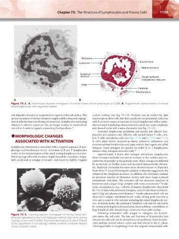
Figure 73–2. A. Transmission electron micrograph of normal human blood lymphocyte (×12,000). B. Diagrammatic representation of normal
blood lymphocyte, with organelles labeled.
and diapedesis localize to nonprotrusive regions of the cell surface. This nuclear outlines (see Fig. 73–1D). Nucleoli may be evident by light
spatial separation of surface receptors might enable a temporal segrega- microscopy in these cells, but they usually are not prominent. Infection
tion of adhesive function during extravasation. Lymphocytes expressing with B. pertussis causes an increase in blood lymphocytes with a partic-
chimeric L-selectin constructs that no longer localize to microvilli do ular activated morphology characterized by small size, scant cytoplasm,
not roll on L-selectin ligands, supporting this hypothesis. 18 and cleaved nuclei with mature chromatin (see Fig. 73–1C).
Activated lymphocytes proliferate and mature into effector lym-
MORPHOLOGIC CHANGES phocytes and memory cells. Effector cells include helper T cells, cyto-
lytic T cells, and plasma cells (see Figs. 73-1F and G, 73–4, and 73–5).
ASSOCIATED WITH ACTIVATION In vitro, plant lectins, bacterial products, polymeric substances, and
enzymes activate lymphocytes and cause mitosis. Such agents are called
Lymphocyte stimulation is associated with a complex sequence of mor- mitogens. Some mitogens are specific for either B or T lymphocytes,
phologic and biochemical events. Activation of B and T lymphocytes whereas other mitogens stimulate both. 19
results in the transformation of the small, resting lymphocyte into pro- Approximately 4 hours after mitogen stimulation, lymphocytes
liferating large cells with abundant highly basophilic cytoplasm, irregu- show increased nucleolar size and an increase in the number and con-
larly condensed or smudgy chromatin, and round to slightly irregular centration of granules in the granular zone. These changes are followed
by an increase in fibrillar zones and increased intranucleolar chroma-
tin. Nucleolar chromatin becomes more electron lucent or dispersed.
From 48 to 72 hours following the addition of phytohemagglutinin, the
volume of the cytoplasm increases. In addition, the cytoplasm contains
an increased number of ribosomal clusters and more rough-surfaced
endoplasmic reticulum. The activated cell has increased numbers of
lysosomes and a larger Golgi complex with more components. Under
20
some circumstances (e.g., cultures of human lymphocytes stimulated
for 7 to 10 days with pokeweed mitogen), some B cells form well-devel-
oped Golgi and plasmacytoid features. Similar plasmacytoid cells are
21
observed in antigen-stimulated lymph nodes, during graft rejection in
vivo, and in some in vitro systems, including the mixed lymphocyte cul-
ture. In lymph nodes, the stimulated lymphoid cells may be referred to
by various pathologists as lymphoblasts, immunoblasts, centroblasts, or
large lymphoid cells. Morphologic criteria for these cells overlap.
Following stimulation with antigen or mitogens, the lympho-
Figure 73–3. Scanning electron micrograph of normal blood lym- cyte enters the cell cycle. The fate and function of lymphocytes that
phocytes separated by the Ficoll-Hypaque method. Cells show varying
numbers of microvilli (×5000). (Used with permission of Dr. Aaron Polliack traverse the cell cycle can be divided into two pathways. Some lympho-
of the Department of Hematology, Hebrew University Hadassah Medical cytes undergo several mitotic cycles and then return to the G phase,
0
School, Jerusalem, Israel.) indistinguishable in morphology from the original nonactivated cells.
Kaushansky_chapter 73_p1135-1148.indd 1139 9/21/15 4:44 PM