
Page 1165 - Williams Hematology ( PDFDrive )
P. 1165
1140 Part IX: Lymphocytes and Plasma Cells
Some of them then become memory cells, programmed to remember
the stimulating antigen and thus respond more rapidly to reexposure
to the original antigen. Alternatively, they become terminally differ-
entiated effector lymphocytes, such as plasma cells or cytotoxic T cells
(Chaps. 75 and 76).
MICROSCOPY AND HISTOCHEMISTRY
OF PLASMA CELLS
MORPHOLOGIC STUDIES
Plasma cells derive from small B lymphocytes after activation in the cor-
rect environment. The characteristic feature of plasma cells is abundant
cytoplasmic and secretory immunoglobulin (Ig). A fully mature plasma
cell lacks surface Ig expression. Each plasma cell has the same clonal rear-
rangement of its V(D)J (variable diversity joining) Ig genes as its predeces-
sor B lymphocyte (Chap. 75). Several mitotic divisions may occur during
cellular differentiation from the resting lymphocyte to the plasmablast to
the immature plasma cell. Immature plasma cells can undergo successive
waves of mitosis in the medullary cords of lymph nodes in response to
antigen. Cell transfer experiments demonstrated that these transformed
22
cells later mature into antibody-producing plasma cells. 23
Pokeweed mitogen induces B lymphocytes to transform into
plasma cells after 7 to 10 days of culture. These plasma cells infre-
24
quently contain large electron-dense inclusions (Russell bodies), which
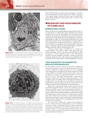
Figure 73–4. Transmission electron micrograph of lymphocyte from 25
20
normal individual incubated with phytohemagglutinin for 3 days. The may measure 2 to 3 μm in diameter (see Fig. 73–1H). Russell bodies,
transformed cell has a large Golgi zone (G) and many ribosomal aggre- cytoplasmic Ig in the endoplasmic reticulum, sometimes are dissolved
gates (arrows). The nucleus is euchromatic (×7500). during the staining procedure. They usually occur in pathologic states
but may be found in plasma cells from normal lymph nodes or marrow.
LIGHT MICROSCOPY, HISTOCHEMISTRY,
AND ELECTRON MICROSCOPY
The mature plasma cell has a characteristic basophilic cytoplasm and
an eccentric nucleus when treated with a polychrome stain. The nuclear
polarity is attributable to a large paranuclear zone, which corresponds
to the Golgi apparatus. The typical mature plasma cell spread on a slide
usually is round or oval and has a diameter of 9 to 20 μm, with a mean
cell diameter of 14 μm and a mean nuclear diameter of 8.5 μm (see
Fig. 73–1F and G). The nuclear heterochromatin is coarse and dis-
26
tributed in a pattern that sometimes resembles the spokes of a wheel
(cartwheel nucleus) on paraffin sections. Normal plasma cells may occa-
sionally have two or more nuclei. Cytochemical features of plasma cells
include positive staining for β-glucuronidase and mitochondrial enzyme
markers. They do not stain for peroxidase or nonspecific esterase. 27
Plasma cells in patients with certain diseases may have different
histochemical properties. Plasma cell size and morphology may be
altered substantially in myeloma and macroglobulinemia (Chaps. 107
and 109, respectively). Plasma cells with two or three nuclei are more
frequent in marrows from patients with plasma cell dyscrasias. Periodic
acid-Schiff stains may reveal cytoplasmic or nuclear inclusions in clonal
plasma cells. Under some circumstances, amyloid inclusions in plasma
28
cells have been detected by electron microscopy. In hemochromatosis
29
and hemosiderosis, plasma cells may contain hemosiderin when exam-
ined by electron microscopy. 30
The plasma cell is packed with a rough-surfaced endoplasmic
Figure 73–5. Transmission electron micrograph of plasmacytoid cell reticulum having numerous attached ribosomes as seen by electron
present in culture of lymphocytes from a patient with chronic lympho- microscopy. A large, circumscribed Golgi zone forms a paranuclear
cytic leukemia incubated with pokeweed mitogen for 7 days. The nucle-
olus (N) and rough-surfaced endoplasmic reticulum (arrows) are evident halo when observed by light microscopy. The nucleus has dense areas
(×9000). (Reproduced with permission from Cohnen G, Douglas SD, Konig E, of heterochromatin. The Golgi zone contains lamellae, vesicles, vacu-
Brittinger G: Pokeweed mitogen response of lymphocytes in chronic lympho- oles, and a number of granules. Mitochondria are located between the
cytic leukemia: A fine structural study, Blood 1973 Oct;42(4):591-600) strands of endoplasmic reticulum. 31
Kaushansky_chapter 73_p1135-1148.indd 1140 9/21/15 4:44 PM