
Page 1166 - Williams Hematology ( PDFDrive )
P. 1166
Chapter 73: The Structure of Lymphocytes and Plasma Cells 1141
ANTIGENS OF HUMAN LYMPHOCYTES lymphoid organs; they are rare in the marrow. These cells appar-
38
ently are enriched for cells that spontaneously produce polyreactive
B LYMPHOCYTE ANTIGENS autoantibodies. 39–41
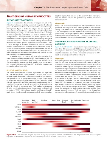
Figure 73–6 summarizes the expression of antigens on cells of the Plasma Cells
B-lymphocyte lineage, including committed progenitor B cells and Many B cell differentiation antigens are not expressed by the mature
pre-B cells. Chapter 74 discusses these cells and the maturation stages plasma cell, including CD20, Pax-5, surface Ig, and HLA class II antigens
they represent. Figure 73–6 also lists antigens that are expressed or (see Fig. 73–6). Of the cells of the B lineage, plasma cells are distinctive
increased upon B-cell activation. Of the B-cell–associated antigens that in that they express CD138 and bright CD38. Clonal plasma cell neo-
42
are commonly used, only a few are restricted to cells of the B lineage. plasms usually have antigen expression distinct from normal plasma cells
Of these antigens, only CD20, CD22, and Pax 5 are not found on other including aberrant expression of CD20, CD28, CD56, and CD117. Clonal
cell types. Pax 5, a transcription factor, is a “master regulator” of B-cell plasma cells usually aberrantly lack expression of CD19 and CD27. 43
development 32,33 that is expressed from the precursor stage through all
B-cell maturation until it is lost at the plasma cell stage. Demonstration
of monoclonal surface Ig allows diagnosis of clonal, neoplastic B cells. T-LYMPHOCYTE AND NATURAL KILLER CELL
CD20 is the target of rituximab, a monoclonal antibody commonly ANTIGENS
used for treatment of B-cell neoplasms. CD19 is restricted mostly to Figure 73–7 and Table 73–1 summarize the expression of antigens on
B cells, but may be expressed weakly by follicular dendritic cells. CD19 cells of the T-lymphocyte and NK lineages. All lymphocyte progeni-
is expressed by B cells at all stages of maturation, including the commit- tors originate in the marrow, but T lymphocytes have their own special
ted B-cell progenitor and most normal plasma cells. As such, it is the organ for maturation—the thymus—whereas NK cells appear to differ-
best-defined pan–B-cell surface antigen. entiate in secondary lymphoid tissue (Chap. 6).
In addition to the CD antigens and Igs, B cells express the three
major histocompatibility complex (MHC) class II antigens: DR, DP, Thymocyte
DQ. These antigens are heterodimers of heavy chains and light chains The thymus promotes the development of antigen-specific T lympho-
that are encoded by genes within the D complex of the human leuko- cytes and eliminates self-reactive T lymphocytes. There are three gen-
cyte antigen (HLA) complex (Chap. 137). MHC class I antigens are eral stages of thymocyte maturation based on the surface CD4 and CD8
expressed on all nucleated cells. expression: double negative, double positive, and single positive. These
stages have corresponding anatomic localization within the thymus with
B-1 B Cells and CD5+ B Cells the least-mature double-negative cells located in the subcapsular area
B-1 B cells have distinctive activation requirements and high levels and the most-mature single-positive cells located in the medulla (Chap.
of CD44 and interleukin (IL)-5 receptor α (IL-5Rα). They prolifer- 6). The most immature T lymphocytes in the thymus populate the sub-
ate more rapidly than other B cells to stimuli such as IgM crosslink- capsular areas and express CD2, CD5, and CD7, antigens present on
ing, possibly because of having constitutively activated nuclear signal T lymphocytes of all stages. Capsular, “double-negative” thymocytes also
transducer and activator of transcription 3 (STAT3). Many, but not all, express CD1a, cytoplasmic CD3, and terminal deoxynucleotidyl trans-
34
B-1 cells express CD5, a 67-kDa transmembrane glycoprotein that is ferase (TdT). The majority of thymocytes are at the “double-positive”
more brightly expressed by T cells. These cells are designated CD5 B stage within the cortical area. These are the cells undergoing positive
cells. B-1 B cells do not express other T-cell markers but do express selection. Once the thymocytes achieve their “education” without
35
all other pan–B-cell surface antigens. Various agents modulate B-cell dying, they mature to the single-positive stage in the medulla. These
expression of CD5. B-1 B cells are found in umbilical cord blood, varying stages of immature T-cell maturation in the thymus have
37
36
adult blood, the pleura and peritoneum, and all major secondary corresponding cell phenotypes in T-lymphoblastic leukemia/lymphoma.
Committed Early Pre-B cell Mature B cell Activated Germinal center Plasma cell
progenitor pre-B cell B cell B cell
CD45
TdT/CD34
CD19
HLA-DR
CD10
CD20/22
Pax-5
clgM
CD38
slgM
clgG/A/E
CD138
Figure 73–6. Clinically useful antigens expressed during B-lymphocyte maturation. The intensity of the antigen expression at each stage of
B-lymphocyte maturation is depicted by gradient density of bars on the graph. c, cytoplasmic; s, surface.
Kaushansky_chapter 73_p1135-1148.indd 1141 9/21/15 4:44 PM