
Page 1287 - Williams Hematology ( PDFDrive )
P. 1287
1262 Part IX: Lymphocytes and Plasma Cells Chapter 82: Mononucleosis Syndromes 1263
27
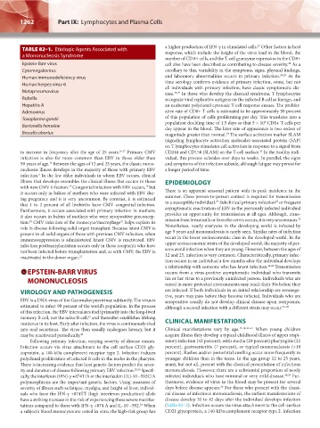
TABLE 82–1. Etiologic Agents Associated with a higher production of IFN-γ in stimulated cells. Other factors in host
response, which include the height of the virus load in the blood, the
a Mononucleosis Syndrome
number of CD 8+ cells, and the T-cell granzyme expression in the CD8+
Epstein-Barr virus cell also have been described as contributing to disease severity. As a
28
Cytomegalovirus corollary to this, variability in the symptoms, signs, physical findings,
Human immunodeficiency virus and laboratory abnormalities occurs in primary infection. 28,29 At the
Human herpes virus-6 time serology confirms evidence of primary infection, some, but not
all individuals with primary infection, have classic symptomatic dis-
Metapneumovirus ease. 28,29 In those who develop the classical syndrome, T lymphocytes
Rubella recognize viral replicative antigens on the infected B cell as foreign, and
Hepatitis A an exuberant polyclonal cytotoxic T-cell response ensues. The prolifer-
Adenovirus ative rate of CD8+ T cells is estimated to be approximately 50 percent
Toxoplasma gondii of this population of cells proliferating per day. This translates into a
9
Bartonella henselae population doubling time of 1.5 days so that 5 × 10 CD8+ T cells per
day appear in the blood. The later rate of appearance is two orders of
Brucella abortus magnitude greater than normal. The surface activation marker SLAM
30
(signaling lymphocyte activation molecule)-associated protein (SAP)
on T lymphocytes stimulates cell activation in response to a signal from
to increase in frequency after the age of 25 years. 16,17 Primary CMV CD244 and CD150 (SLAM) on the T-cell surface. In the healthy indi-
31
infection is also far more common than EBV in those older than vidual, this process subsides over days to weeks. In parallel, the signs
16
50 years of age. Between the ages of 12 and 25 years, the classic mono- and symptoms of the infection subside, although fatigue may persist for
nucleosis illness develops in the majority of those with primary EBV a longer period of time.
infection. In the few older individuals in whom EBV occurs, clinical
8
illness that develops resembles the clinical illness that occurs in those EPIDEMIOLOGY
18
with new CMV infection. Congenital infection with EBV occurs, but
19
it occurs only in babies of mothers who were infected with EBV dur- There is an apparent seasonal pattern with its peak incidence in the
ing pregnancy and it is very uncommon. By contrast, it is estimated summer. Close person-to-person contact is required for transmission
29
28
that 1 to 2 percent of all livebirths have CMV congenital infection. to a susceptible individual. Subclinical primary infection or frequent
Furthermore, it occurs associated with primary infection in mothers; asymptomatic reactivation of EBV in the previously infected individual
it also occurs in babies of mothers who were seropositive preconcep- provides an opportunity for transmission at all ages. Although, trans-
32
20
21
tion. CMV infection of the monocyte/macrophage helps explain its mission from breast milk or from the cervix occurs, it is very uncommon.
role in disease following solid-organ transplant. Because latent CMV is Nonetheless, nearly everyone in the developing world is infected by
present in all solid organs of those with previous CMV infection, when age 5 years and mononucleosis is rarely seen. Similar rates of infection
immunosuppression is administered latent CMV is reactivated. EBV occur in the lower socioeconomic class in the developed world. In the
infection posttransplantation occurs only in those recipients who have upper socioeconomic strata of the developed world, the majority of per-
not been infected before transplantation and, as with CMV, the EBV is sons avoid infection when they are young. However, between the ages of
reactivated in the donor organ. 22 12 and 25, infection is very common. Characteristically, primary infec-
tion occurs in an individual a few months after the individual develops
a relationship with someone who has latent infection. 24,28 Transmission
EPSTEIN-BARR VIRUS occurs from a virus-positive asymptomatic individual who transmits
MONONUCLEOSIS his or her virus to a previously uninfected person. Individuals who are
raised in more protected environments may reach their 30s before they
VIROLOGY AND PATHOGENESIS are infected. If both individuals in an initial relationship are seronega-
tive, years may pass before they become infected. Individuals who are
EBV is a DNA virus of the Gammaherpesvirinae subfamily. The virus is seropositive usually do not develop clinical disease upon reexposure,
estimated to infect 90 percent of the world’s population. In the process although a second infection with a different strain may occur. 33–35
of this infection, the EBV intercalates itself primarily into the long-lived
memory B cell, not the naïve B cell, and thereafter establishes lifelong
23
residence in its host. Early after infection, the virus is continuously shed CLINICAL MANIFESTATIONS
into oral secretions. The virus then usually undergoes latency, but it Clinical manifestations vary by age. 15,18,36–41 When young children
may be reactivated periodically. 24 acquire illness they develop a typical childhood illness of upper respi-
Following primary infection, varying severity of disease ensues. ratory infection (43 percent), otitis media (29 percent) pharyngitis (21
Infection occurs via virus attachment to the cell surface CD21 gly- percent), gastroenteritis (7 percent), or typical mononucleosis (<10
coprotein, a 140-kDa complement receptor type 2. Infection induces percent). Rashes and/or periorbital swelling occur more frequently in
polyclonal proliferation of infected B cells in the nodes in the pharynx. younger children than in the teens. In the age group 12 to 25 years,
There is increasing evidence that host genetic factors predict the sever- many, but not all, present with the classical presentation of infectious
ity and duration of disease following primary EBV infection. 25,26 Specifi- mononucleosis. However, there are a substantial proportion of newly
cally, the interferon (IFN)-γ +874T/A or the interleukin (IL)-10 –592C/A infected individuals who have minimal or very mild disease. 28,29 Fur-
polymorphisms are the important genetic factors. Using measures of thermore, evidence of virus in the blood may be present for several
severity of illness such as fatigue, myalgia, and height of fever, individ- days before disease appears. For those who present with the classi-
28
uals who have the IFN-γ +874TT (high interferon production) allele cal disease of infectious mononucleosis, the earliest manifestations of
have a striking increase in the risk of experiencing these severe manifes- disease develop 35 to 42 days after the individual develops infection
tations compared to those with IFN-γ +874 A and IL-10 –592. When (Table 82–2). Infection occurs via virus attachment to the cell-surface
24
a subject’s blood monocytes are tested in vitro, the high-risk group has CD21 glycoprotein, a 140-kDa complement receptor type 2. Infection
Kaushansky_chapter 82_p1261-1272.indd 1262 9/18/15 10:04 AM