
Page 1289 - Williams Hematology ( PDFDrive )
P. 1289
1264 Part IX: Lymphocytes and Plasma Cells Chapter 82: Mononucleosis Syndromes 1265
LABORATORY FINDINGS not appear until the recovery phase of the illness. For those patients
Antibody Responses suspected of having infectious mononucleosis but who do not develop
heterophile antibody, detection of IgG and IgM antibody to VCA with
By week three of a mononucleosis caused by primary EBV infection, an the absence of EBNA-1 antibody leads to the diagnosis. A real-time
49
heterophile antibody response will occur in approximately 85 percent of positive polymerase chain reaction (PCR) is sometimes useful. 50
patients. The test is called the monospot test and may be falsely negative,
especially in young children. 36,48 Reactive Lymphocytosis
The infection of B cells results in their production of a variety of anti- Expansion of cytotoxic T lymphocytes produces lymphocytosis. Reac-
bodies against uninvolved infectious agents. The B-cell clones expanded, tive lymphocytes are larger than the lymphocytes normally found in the
non-specifically, result in antibodies against Chlamydia, Borrelia burg- blood (Fig. 82–1). They may have a vacuolated cytoplasm, lobulated and
dorferi, the yellow fever virus, and many other infectious agents. If the eccentrically placed nucleus, and a cell membrane that often is indented
patient’s febrile illness is considered a fever of unknown origin, diagnos- by neighboring erythrocytes. A more darkly staining peripheral cyto-
tic conclusions may be misleading. A variety of other antibodies against plasm, called “skirting,” occurs. Reactive lymphocytes are a hematologic
antigens that are not those of infectious agents also are produced because hallmark of infectious mononucleosis, but they are not always found
29
of polyclonal B-cell activation. These include antiplatelet, anti–red cell and are not pathognomonic. They also are found in CMV infection,
(anti-i cold agglutinin), and antinuclear antibodies. roseola (caused by human herpes virus-6), viral hepatitis, toxoplasmo-
At the time clinical disease is evident, both immunoglobulin (Ig) sis, rubella, mumps, and drug reactions.
G and IgM antibodies to Epstein-Barr virus capsid antigen (VCA) usu- Sheets of lymphocytes are noted on a stained slide preparation
ally are detectable. Later, antibody to early antigen (EA) develops. A of tonsillar exudate. The immunophenotype of lymphocytes in mono-
small proportion of individuals will also have developed antibody to nucleosis syndromes assessed by multiparametric flow cytometry has
EBNA-1 on presentation. However, usually EBNA-1 antibody does confirmed that lymphocytosis results from CD8+ T cells. CD4+ T
A B
C D
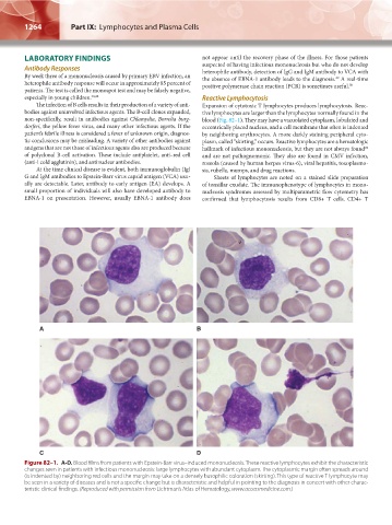
Figure 82–1. A-D. Blood films from patients with Epstein-Barr virus–induced mononucleosis. These reactive lymphocytes exhibit the characteristic
changes seen in patients with infectious mononucleosis: large lymphocytes with abundant cytoplasm. The cytoplasmic margin often spreads around
(is indented by) neighboring red cells and the margin may take on a densely basophilic coloration (skirting). This type of reactive T lymphocyte may
be seen in a variety of diseases and is not a specific change but is characteristic and helpful in pointing to the diagnosis in concert with other charac-
teristic clinical findings. (Reproduced with permission from Lichtman’s Atlas of Hematology, www.accessmedicine.com.)
Kaushansky_chapter 82_p1261-1272.indd 1264 9/18/15 10:05 AM