
Page 1303 - Williams Hematology ( PDFDrive )
P. 1303
1278 Part X: Malignant Myeloid Diseases Chapter 83: Classification and Clinical Manifestations of the Clonal Myeloid Disorders 1279
aneuploid or pseudodiploid cells in the clone, are the result of the expan- MODERATE-DEVIATION CLONAL
sion of a somatically mutated cell, and can be associated with significant MYELOID DISORDERS
morbidity and premature death; thus, they are neoplasias not dysplasias.
They demonstrate clonal (genomic) instability, and each has a propen- Primary myelofibrosis (Chap. 86) and CML (Chap. 89) classically
sity to evolve into AML at a rate that far exceeds the incidence of the dis- share the features of overproduction of granulocytes and platelets and
ease in the general population. The term myelodysplasia was proposed impaired production of red cells. In contrast to the minimally devi-
at a conference in Paris in 1976 at a time when prominent dysmorpho- ated clonal myeloid neoplasms, CML and primary myelofibrosis may
genesis and cytopenias were thought to be the singular abnormalities have a small to moderate proportion of leukemic blast cells in marrow
and arguments existed as to whether some of these syndromes without and blood. The most constant feature in primary myelofibrosis is the
increased blast cell percentages represented a preneoplastic (preleuke- abundance of neoplastic, dysmorphic megakaryocytes and the resul-
mic) condition. They have long been established as neoplastic (a spec- tant predisposition to marrow reticulin and collagen fibrosis, osteo-
11
trum of minimal-deviation to severe-deviation leukemias)—indeed, sclerosis, extramedullary fibrohematopoietic tumors, splenomegaly,
those with overt leukemic hematopoiesis (quantitatively increased leu- and teardrop-shaped red cells (dacryocytes) in every oil immersion
kemic blast cell counts), which made up approximately 50 percent of field on the blood film. The megakaryocytic abnormalities are so domi-
cases, were known at the time to be neoplasms—but the terminology nant and consistent in this disorder that it could be considered chronic
19
has not been rectified. megakaryocytic leukemia. The cells in this disorder have no specific
cytogenetic change, but approximately 50 percent of cases carry a muta-
OVERPRODUCTION OF CELLS PROMINENT tion in the JAK2 gene and approximately one-third have wild-type JAK2
but a mutation in the CALR gene (Chap. 86).
13,14
These two mutations
Polycythemia vera (Chap. 84) and essential thrombocythemia (Chap. 85) give primary myelofibrosis a genetic kinship with polycythemia vera
are clonal myeloid disorders so named because of the overaccumulation and essential thrombocytosis. They are often referred to as “the mye-
of red cells, and often neutrophils, and platelets in the blood in poly- loproliferative neoplasms,” but virtually all clonal myeloid diseases are
cythemia, and of platelets, and to a lesser extent neutrophils, in throm- fundamentally myeloproliferative as the term refers, principally, to mar-
bocythemia. Each cell lineage is affected in each disorder, reflecting a row hematopoiesis. The clinical behavior of primary myelofibrosis is,
12
multipotential hematopoietic cell origin, but the magnitude of the effect in most cases of a progressive neoplasm with morphologic evidence,
on each lineage differs. The decrease in red cell production in essential of lower-level leukemic hematopoiesis and with a median survival sig-
thrombocythemia usually is slight to mild. Polycythemia vera and essen- nificantly less than polycythemia vera or essential thrombocythemia.
tial thrombocythemia do not show morphologic evidence of leuke- Primary myelofibrosis is another misnomer perpetuated in the WHO
mic hematopoiesis; the proportion of blast cells in the marrow is never classification. The fibrosis is secondary to cytokines released by neoplas-
increased above normal, and blast cells are never present in the blood. tic (leukemic) megakaryocytes (an epiphenomenon) and it is the only
Hematopoietic differentiation and maturation are maintained. These are cancer in the medical lexicon named after connective tissue fibers and
minimal-deviation neoplasms. These disorders do not have a specific not the cells in which the cancer arises. 19
cytogenetic abnormality, but approximately 95 percent of cases of poly- In contrast to primary myelofibrosis, CML has a rearrangement of
cythemia and approximately 50 percent of cases of essential thrombo- the breakpoint cluster (BCR) gene on chromosome 22. The shortening
cythemia have an acquired mutation in the Janus kinase 2 (JAK2) gene. In of the long arm of chromosome 22 gives it the designation of the Phil-
thrombocythemia, 25 percent of patients have wild-type JAK2 genes and adelphia chromosome, now called the Ph chromosome. It can be iden-
mutations in the calreticulin (CALR) gene. A few percent of patients with tified by Giemsa (G)-banding cytogenetic studies in approximately 90
thrombocythemia have nonmutated JAK2 and CALR but a mutation in percent of patients with CML. This mutation is caused by and is a reflec-
the myeloproliferative leukemia virus gene (MPL; Chaps. 84 and 85). 13,14 tion of the translocation t(9;22)(q34;q11)(BCR-ABL1 [Abelson murine
Several studies of comparative survival of the chronic myeloproliferative leukemia viral oncogene homologue 1]). The BCR-ABL1 fusion in CML
neoplasms have been reported. 4,15–18,18a In the most comprehensive study cells can be found in virtually all cases studied by fluorescence in situ
of survival as of this writing, patients with essential thrombocythemia hybridization or the polymerase chain reaction. Only approximately 4
have only slightly decreased survival than expected over 10 years of obser- percent of patients with a phenotype indistinguishable from BCR-re-
vation, but this widens somewhat over longer periods. The difference in arrangement–positive CML do not have the rearrangement (see Table
survival of patients with primary myelofibrosis is dramatically less than 83-1 and Chap. 89). An unrelenting increase in the white cell (granulo-
expected for age- and gender-matched unaffected persons and the sur- cyte) count, anemia, splenomegaly, and a progressive course are com-
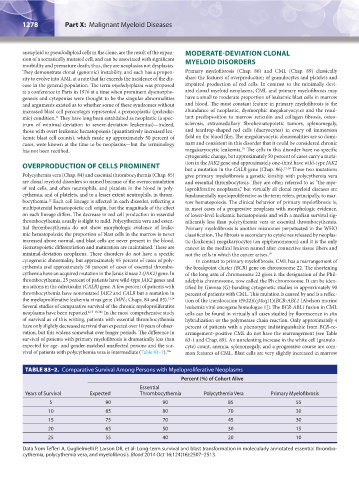
vival of patients with polycythemia vera is intermediate (Table 83–2). 18a mon features of CML. Blast cells are very slightly increased in marrow
TABLE 83–2. Comparative Survival Among Persons with Myeloproliferative Neoplasms
Percent (%) of Cohort Alive
Essential
Years of Survival Expected Thrombocythemia Polycythemia Vera Primary Myelofibrosis
5 90 90 85 55
10 85 80 70 30
15 75 70 45 30
20 65 50 30 15
25 55 40 20 10
Data from Tefferi A, Guglielmelli P, Larson DR, et al: Long-term survival and blast transformation in molecularly annotated essential thrombo-
cythemia, polycythemia vera, and myelofibrosis. Blood 2014 Oct 16;124(16):2507–2513.
Kaushansky_chapter 83_p1273-1290.indd 1278 9/21/15 11:12 AM