
Page 1308 - Williams Hematology ( PDFDrive )
P. 1308
1282 Part X: Malignant Myeloid Diseases Chapter 83: Classification and Clinical Manifestations of the Clonal Myeloid Disorders 1283
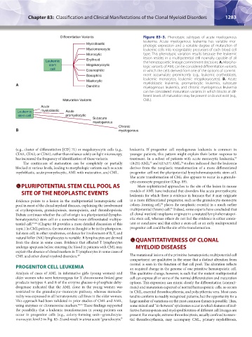
Differentiation Variants Figure 83–3. Phenotypic subtypes of acute myelogenous
leukemia. Acute myelogenous leukemia has variable mor-
Myeloblastic phologic expression and a variable degree of maturation of
Myelomonocytic leukemic cells into recognizable precursors of each blood cell
Monocytic type. This phenotypic variation results because the leukemic
Leukemic Erythroid lesion resides in a multipotential cell normally capable of all
the hematopoietic lineage commitment decisions. A. Morpho-
stem Megakaryocytic logic variants of AML can be considered differentiation variants
cell Eosinophilic in which the cells derived from one of the options of commit-
Basophilic ment accumulate prominently (e.g., leukemic erythroblasts,
Mastocytic leukemic monocytes, leukemic megakaryocytes). B. Acute
A Dendritic myeloblastic leukemia, promyelocytic leukemia, subacute
myelogenous leukemia, and chronic myelogenous leukemia
can be considered maturation variants in which blocks at dif-
ferent levels of maturation may be present or do not exist (e.g.,
Maturation Variants CML).
Acute
Leukemic myeloblastic Acute
stem cell promyelocytic
Subacute
myelogenous
Chronic
B myelogenous
(e.g., cluster of differentiation [CD] 71) or megakaryocytic cells (e.g., leukemia. If progenitor cell myelogenous leukemia is common in
CD41, CD42, or CD61), rather than reliance solely on light microscopy, younger patients, this pattern might explain their better response to
52
has increased the frequency of identification of these variants. treatment. In a subset of patients with acute monocytic leukemia,
The continuum of maturation can be completely or partially t(8;21) AML, and t(15;17) AML, studies indicated that the leukemia
53
54
blocked at various levels, leading to morphologic variants such as acute derives from the neoplastic transformation of a more differentiated
myeloblastic, acute promyelocytic, AML with maturation, and CML. progenitor cell not the pluripotential lymphohematopoietic stem cell.
The acute transformation of CML also appears to occur in a granulo-
cyte-monocyte progenitor (Chap. 89).
PLURIPOTENTIAL STEM CELL POOL AS More sophisticated approaches to the site of the lesion in mouse
SITE OF THE NEOPLASTIC EVENTS models of AML have indicated that disorders like acute promyelocytic
leukemia for which there is evidence in humans that it may originate
Evidence points to a lesion in the multipotential hematopoietic cell in a more differentiated progenitor, such as the granulocyte-monocyte
54
pool in most of the clonal myeloid diseases, explaining the involvement colony-forming cell, places the neoplastic event(s) in a much earlier
55
of erythropoiesis, granulopoiesis, monopoiesis, and thrombopoiesis. multipotential (?stem) cell. Indeed, some experts have concluded that
Debate continues whether the cell of origin is a pluripotential (lympho- all clonal myeloid neoplasms originate in a mutated lymphohematopoi-
hematopoietic) stem cell or a somewhat more differentiated multipo- etic stem cell, whereas others do not feel the evidence is either consis-
tential cell. 47,48 (Chapter 88 provides a more detailed discussion of this tent or conclusive and that either a stem cell or an early multipotential
topic.) In CML patients, the mutation is thought to be in the pluripoten- progenitor cell could be the site of the transformation.
tial stem cell; in other syndromes, evidence for involvement of B, T, and
natural killer (NK) lymphocytes is variable. B lymphocytes are derived QUANTITATIVENESS OF CLONAL
from the clone in some cases. Evidence that affected T lymphocytes
undergo apoptosis before entering the blood in patients with CML may MYELOID DISEASES
explain the absence of clonal markers in T lymphocytes in some cases of
CML and other clonal myeloid disorders. 49 The mutational lesions of the primitive hematopoietic multipotential cell
compartment are qualitative in the sense that a distinct alteration from
PROGENITOR CELL LEUKEMIA normal is seen in the function of that cell pool. The alteration reflects
an acquired change in the genome of one primitive hematopoietic cell.
Analysis of cases of AML in informative girls (young women) and This qualitative change, however, is such that the mutant multipotential
older women who were heterozygous for X chromosome-linked gene cell can express all or some of the normal differentiation and maturation
products isotypes A and B of the enzyme glucose-6-phosphate dehy- options. This expression can mimic closely the differentiation (commit-
drogenase indicated that the AML clone in the young women was ment) and maturation expected of normal hematopoietic cells, as occurs
restricted to the granulocyte–monocyte pathway, whereas monoclo- in CML, essential thrombocythemia, and polycythemia vera. Most cases
nality was expressed in all hematopoietic cell lines in the older women. tend to conform to readily recognized patterns, but the opportunity for a
This approach had been validated in prior studies of CML and AML, large number of variations on the most common themes is possible. Thus,
using enzymes or chromosome markers. 50,51 These findings supported some mixed and “in-between” syndromes occur in which features of inef-
the possibility that a leukemic transformation in young patients can fective hematopoiesis and myeloproliferation of different cell lineages are
occur in progenitor cells (e.g., colony-forming unit—granulocyte- present. For example, extreme thrombocytosis, usually confined to essen-
monocyte; level 3 in Fig. 83–2) and result in a true acute “granulocytic” tial thrombocythemia, may accompany CML, primary myelofibrosis,
Kaushansky_chapter 83_p1273-1290.indd 1283 9/21/15 11:13 AM