
Page 1307 - Williams Hematology ( PDFDrive )
P. 1307
1282 Part X: Malignant Myeloid Diseases Chapter 83: Classification and Clinical Manifestations of the Clonal Myeloid Disorders 1283
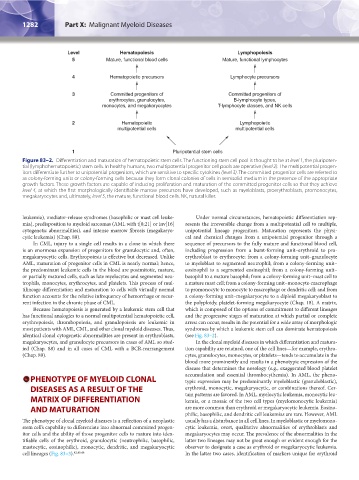
Level Hematopoiesis Lymphopoiesis
5 Mature, functional blood cells Mature, functional lymphocytes
4 Hematopoietic precursors Lymphocyte precursors
3 Committed progenitors of Committed progenitors of
erythrocytes, granulocytes, B-lymphocyte types,
monocytes, and megakaryocytes T-lymphocyte classes, and NK cells
2 Hematopoietic Lymphopoietic
multipotential cells multipotential cells
1 Pluripotential stem cells
Figure 83–2. Differentiation and maturation of hematopoietic stem cells. The functioning stem cell pool is thought to be at level 1, the pluripoten-
tial (lymphohematopoietic) stem cells. In healthy humans, two multipotential progenitor cell pools are operative (level 2). The multipotential progen-
itors differentiate further to unipotential progenitors, which are sensitive to specific cytokines (level 3). The committed progenitor cells are referred to
as colony-forming units or colony-forming cells because they form clonal colonies of cells in semisolid medium in the presence of the appropriate
growth factors. These growth factors are capable of inducing proliferation and maturation of the committed progenitor cells so that they achieve
level 4, at which the first morphologically identifiable marrow precursors have developed, such as myeloblasts, proerythroblasts, promonocytes,
megakaryocytes and, ultimately, level 5, the mature, functional blood cells. NK, natural killer.
leukemia), mediator-release syndromes (basophilic or mast cell leuke- Under normal circumstances, hematopoietic differentiation rep-
mia), predisposition to myeloid sarcomas (AML with t[8;21] or inv[16] resents the irreversible change from a multipotential cell to multiple,
cytogenetic abnormalities), and intense marrow fibrosis (megakaryo- unipotential lineage progenitors. Maturation represents the physi-
cytic leukemia) (Chap. 88). cal and chemical changes from a unipotential progenitor through a
In CML, injury to a single cell results in a clone in which there sequence of precursors to the fully mature and functional blood cell,
is an enormous expansion of progenitors for granulocytic and, often, including progression from a burst-forming unit–erythroid to pro-
megakaryocytic cells. Erythropoiesis is effective but decreased. Unlike erythroblast to erythrocyte; from a colony-forming unit–granulocyte
AML, maturation of progenitor cells in CML is nearly normal; hence, to myeloblast to segmented neutrophil; from a colony-forming unit–
the predominant leukemic cells in the blood are postmitotic, mature, eosinophil to a segmented eosinophil; from a colony-forming unit–
or partially matured cells, such as late myelocytes and segmented neu- basophil to a mature basophil; from a colony-forming unit–mast cell to
trophils, monocytes, erythrocytes, and platelets. This process of mul- a mature mast cell; from a colony-forming unit–monocyte-macrophage
tilineage differentiation and maturation to cells with virtually normal to promonocyte to monocyte to macrophage or dendritic cell; and from
function accounts for the relative infrequency of hemorrhage or recur- a colony-forming unit–megakaryocyte to a diploid megakaryoblast to
rent infection in the chronic phase of CML. the polyploidy, platelet-forming megakaryocyte (Chap. 18). A matrix,
Because hematopoiesis is generated by a leukemic stem cell that which is composed of the options of commitment to different lineages
has functional analogies to a normal multipotential hematopoietic cell, and the progressive stages of maturation at which partial or complete
erythropoiesis, thrombopoiesis, and granulopoiesis are leukemic in arrest can occur, results in the potential for a wide array of morphologic
most patients with AML, CML, and other clonal myeloid diseases. Thus, syndromes by which a leukemic stem cell can dominate hematopoiesis
identical clonal cytogenetic abnormalities are present in erythroblasts, (see Fig. 83–2).
megakaryocytes, and granulocyte precursors in cases of AML so stud- In the clonal myeloid diseases in which differentiation and matura-
ied (Chap. 88) and in all cases of CML with a BCR-rearrangement tion capability are retained, one of the cell lines—for example, erythro-
(Chap. 89). cytes, granulocytes, monocytes, or platelets—tends to accumulate in the
blood more prominently and results in a phenotypic expression of the
disease that determines the nosology (e.g., exaggerated blood platelet
PHENOTYPE OF MYELOID CLONAL accumulation and essential thrombocythemia). In AML, the pheno-
typic expression may be predominantly myeloblastic (granuloblastic),
DISEASES AS A RESULT OF THE erythroid, monocytic, megakaryocytic, or combinations thereof. Cer-
MATRIX OF DIFFERENTIATION tain patterns are favored. In AML, myelocytic leukemia, monocytic leu-
kemia, or a mosaic of the two cell types (myelomonocytic leukemia)
AND MATURATION are more common than erythroid or megakaryocytic leukemia. Eosino-
philic, basophilic, and dendritic cell leukemias are rare. However, AML
The phenotype of clonal myeloid diseases is a reflection of a neoplastic usually has a disturbance in all cell lines. In myeloblastic or myelomono-
stem cell’s capability to differentiate into abnormal committed progen- cytic leukemia, overt, qualitative abnormalities of erythroblasts and
itor cells and the ability of those progenitor cells to mature into iden- megakaryocytes may occur. The prevalence of the abnormalities in the
tifiable cells of the erythroid, granulocytic (neutrophilic, basophilic, latter two lineages may not be great enough or evident enough for the
mastocytic, eosinophilic), monocytic, dendritic, and megakaryocytic observer to designate a case as erythroid or megakaryocytic leukemia.
cell lineages (Fig. 83–3). 42,45,46 In the latter two cases, identification of markers unique for erythroid
Kaushansky_chapter 83_p1273-1290.indd 1282 9/21/15 11:13 AM