
Page 1306 - Williams Hematology ( PDFDrive )
P. 1306
1280 Part X: Malignant Myeloid Diseases Chapter 83: Classification and Clinical Manifestations of the Clonal Myeloid Disorders 1281
thrombocytopenia, among other changes (Chap. 89). The progression PATHOGENESIS OF CLONAL
from chronic to accelerated phase or blast phase of CML, however, has
been delayed in the majority of patients by the application of tyrosine MYELOID DISEASES
kinase inhibitor therapy during the chronic phase of the disease. Deter-
mining the frequency of evolution to AML in those patients with CML In AML, a sequence of mutations in a single multipotential cell results
who enter a complete molecular remission with tyrosine kinase inhibi- in a clone that is severely defective and contains precursor cells that are
42,43
tors must await observations over the next decade. largely unable to mature. Proliferation of primitive progenitors is
This process of clonal evolution is an intrinsic feature of the excessive when considered in absolute terms, that is, the total number
genomic instability of clonal myeloid diseases. The practice of calling of blast cells proliferating. AML is a clinical disease with many forms of
the result of this process “secondary AML” is obfuscating. This choice of morphologic expression. This variation of phenotype is consistent with
terms is notable in the case of myelodysplastic syndrome, which is “leu- the large number of genetic lesions identified and the behavior of the
kemia” at the time of diagnosis. (Leukemia is defined as the neoplastic leukemic stem cell, which is capable of differentiation into all the blood
transformation of a primitive multipotential hematopoietic [myeloid] cell lineages (Fig. 83–1). Hence, the asymmetrical and uncoordinated
cell.) The neoplastic transformation has occurred and the progression differentiation and maturation of leukemic progenitor cells may allow
44
to a more advanced myeloid neoplasm is a process quite different from one or another cell type to predominate. The different morphologic
the secondary AML that occurs as a result of recent chemotherapy for or cytogenetic variants of AML are each rapidly progressive, however, if
a lymphoma or an unrelated cancer (e.g., breast cancer). When there is not treated successfully (Chap. 88).
progression to AML from a previously diagnosed clonal myeloid dis- Important epiphenomena are related to certain morphologic types
ease, it should be designated as clonally evolved AML (ceAML). This of AML, such as tissue infiltration, including into the central nervous
distinction is important because an effort to develop methods to prevent system in monocytic leukemia, disseminated intravascular coagula-
clonal evolution is very likely to be different from methods to prevent tion, fibrinolysis, and hemorrhage in promyelocytic leukemia, and to a
true secondary leukemia. lesser extent in monocytic leukemia, hepatosplenomegaly (eosinophilic
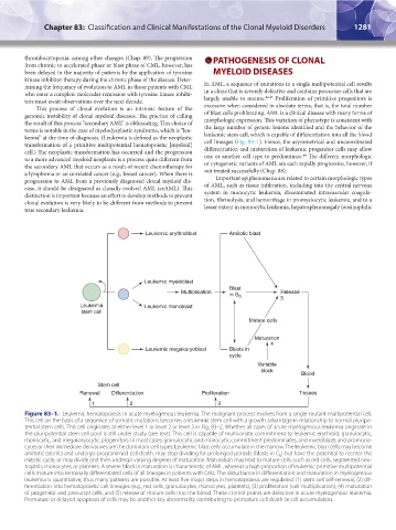
Leukemic erythroblast Amitotic blast
Leukemic myeloblast
Blast
Multiplication in G 0 Release
5
Leukemia Leukemic monoblast
stem cell
Mature cells
Maturation
4
Leukemic megakaryoblast Blasts in
cycle
Variable
block
Blood
Stem cell
Renewal Differentiation Proliferation Tissues
1 2 3
Figure 83–1. Leukemic hematopoiesis in acute myelogenous leukemia. The malignant process evolves from a single mutant multipotential cell.
This cell on the basis of a sequence of somatic mutations becomes a leukemia stem cell with a growth advantage in relationship to normal pluripo-
tential stem cells. This cell originates at either level 1 or level 2 or level 3 in Fig. 83–2. Whether all cases of acute myelogenous leukemia originate in
the pluripotential stem cell pool is still under study (see text). This cell is capable of multivariate commitment to leukemic erythroid, granulocytic,
monocytic, and megakaryocytic progenitors. In most cases, granulocytic and monocytic commitment predominates, and myeloblasts and promono-
cytes or their immediate derivatives are the dominant cell types. Leukemic blast cells accumulate in the marrow. The leukemic blast cells may become
amitotic (sterile) and undergo programmed cell death, may stop dividing for prolonged periods (blasts in G ) but have the potential to reenter the
0
mitotic cycle, or may divide and then undergo varying degrees of maturation. Maturation may lead to mature cells, such as red cells, segmented neu-
trophils, monocytes, or platelets. A severe block in maturation is characteristic of AML, whereas a high proportion of leukemic primitive multipotential
cells mature into terminally differentiated cells of all lineages in patients with CML. The disturbance in differentiation and maturation in myelogenous
leukemia is quantitative, thus many patterns are possible. At least five major steps in hematopoiesis are regulated: (1) stem cell self-renewal, (2) dif-
ferentiation into hematopoietic cell lineages (e.g., red cells, granulocytes, monocytes, platelets), (3) proliferation (cell multiplication), (4) maturation
of progenitor and precursor cells, and (5) release of mature cells into the blood. These control points are defective in acute myelogenous leukemia.
Premature or delayed apoptosis of cells may be another key abnormality contributing to premature cell death or cell accumulation.
Kaushansky_chapter 83_p1273-1290.indd 1281 9/21/15 11:13 AM