
Page 1309 - Williams Hematology ( PDFDrive )
P. 1309
1284 Part X: Malignant Myeloid Diseases Chapter 83: Classification and Clinical Manifestations of the Clonal Myeloid Disorders 1285
or clonal bicytopenia with thrombocytosis. Erythrocytosis may rarely Normal cells reduced
accompany CML. Atypical myeloproliferative syndromes or other clonal Normal or unapparent
myeloid diseases may have mixtures of anemia, granulocytopenia, and hematopoiesis
thrombocytosis or of anemia, granulocytosis, and thrombocytopenia
rather than pancytopenia. Qualitative abnormalities of red cell, granu- Blocked by leukemic cells
locyte, or platelet structure or function may be more or less prominent
in a given patient. For example, qualitative abnormalities of erythroblast Leukemic cells predominate
development may result in acquired α-thalassemia (acquired hemo- Leukemic (~1 trillion cells)
globin H disease), especially in patients with primary myelofibrosis, or hematopoiesis
occasionally other clonal myeloid diseases. In AML, unusual patterns of A
phenotypic expression occur frequently. For example, prominent leuke-
mic erythroblasts and monocytes or eosinophils and monocytes may be Normal cells
seen in patients. So much opportunity for variation in disease expression Normal markedly reduced or unapparent
exists among patients with AML that observation of patients in whom the hematopoiesis
phenotype of their leukemic cells is identical to the phenotype of other
patients is unusual. Choice of treatment is little affected by these varia- Blocked by cytotoxic drugs
tions. Decisions about whether to treat and which drugs to use are greatly
influenced by whether a patient has a chronic, subacute, or acute clonal
myeloid disease; by the rate of progression of the disease; by the extent of Leukemic cells unapparent
Leukemic
the leukemic blast cell infiltrate; by the cytogenetic findings; and by the hematopoiesis (<1 billion cells)
severity of the cytopenias. The experienced diagnostician and therapist
usually can identify variants as a clonal myeloid disorder and can manage Latrogenic aplastic pancytopenia provides opportunity
the disorder as dictated by their manifestations regardless of their precise for reemergence of normal hematopoiesis
subclassification. B
Normal cells
INTERPLAY OF CLONAL AND repopulate marrow and blood
POLYCLONAL HEMATOPOIESIS Normal
hematopoiesis
Although potentially curative chemotherapy of myelogenous leuke- Blocked
mia was introduced in the mid-20th century to kill “the last leukemic
cell,” two important factors were not explicitly appreciated. The first
was whether residual normal stem cells coexisted in marrow to restore Leukemic
polyclonal (normal) hematopoiesis if ablation of the leukemia was hematopoiesis Leukemic cells unapparent
accomplished. The second was whether, given the estimates of 1 trillion C
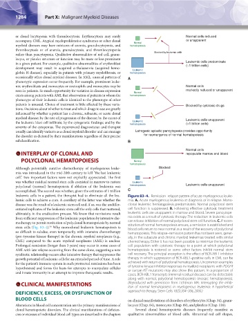
leukemic cells in a patient, the therapist had to eliminate all the leu- Figure 83–4. Remission–relapse pattern of acute myelogenous leuke-
kemic cells to achieve a cure. A corollary of the latter was whether the mia. A. Acute myelogenous leukemia at diagnosis or in relapse. Mono-
disease was the result of a leukemic stem cell and, if so, was the undiffer- clonal leukemic hematopoiesis predominates. Normal polyclonal stem
entiated replicates of the leukemic stem cell the only cells that mattered, cell function is suppressed. B. Following effective cytotoxic treatment
ultimately, in the eradication process. We know that remissions result leukemic cells are unapparent in marrow and blood. Severe pancytope-
from sufficient suppression of the leukemic population by intensive che- nia exists as a result of cytotoxic therapy. The reduction in leukemic cells
motherapy to permit restitution of polyclonal hematopoiesis by normal can release inhibition of normal polyclonal stem cell function. C. If recon-
stem cells (Fig. 83–4). Why monoclonal leukemic hematopoiesis is stitution of normal hematopoiesis ensues, a remission is established and
56
blood cells return to near normal as a result of the recovery of polyclonal
so difficult to subdue, even temporarily, with intensive chemotherapy hematopoiesis. This relapse–remission pattern has not been seen, gener-
(pre–tyrosine kinase therapy) in the chronic myeloid neoplasms (e.g., ally, in the subacute and chronic myeloid leukemias treated with similar
CML) compared to the acute myeloid neoplasms (AML) is unclear. chemotherapy. Either it has not been possible to minimize the leukemic
Prolonged remission (longer than 3 years) may occur in some cases of cell population with cytotoxic therapy to a point at which polyclonal
AML with late relapse occurring from the same clone, suggesting a new hematopoiesis is restored or some other factors inhibit normal stem
symbiotic relationship occurs after intensive therapy that suppresses the cell recovery. The principal exception is the effect of BCR-ABL1 inhibitor
growth potential of leukemic cells for an extended period of time. A role therapy in which suppression of BCR-ABL1–positive cells in CML can be
for the patient’s immune system in such protracted remissions has been achieved with return of polyclonal hematopoiesis. Uncommon examples
hypothesized and forms the basis for attempts to manipulate cellular of tyrosine kinase inhibitor responses in myeloid neoplasms with PDGFR
and innate immunity in an attempt to improve therapeutic results. or certain KIT mutations may also show this pattern. In a proportion of
cases, BCR-ABL1 transcripts (minimal residual disease) can be detectable
along with normal, polyclonal hematopoiesis (mosaic hematopoiesis).
CLINICAL MANIFESTATIONS (Reproduced with permission from Lichtman MA: Interrupting the inhib-
iton of normal hematopoiesis in myelogenous leukemia: A hypothetical
DEFICIENCY, EXCESS, OR DYSFUNCTION OF approach to therapy. Stem Cells 18(5):304–306, 2000.)
BLOOD CELLS on clinical manifestations of disorders of erythrocytes (Chap. 34), granu-
Alterations in blood cell concentration are the primary manifestations of locytes (Chap. 64), monocytes (Chap. 69), and platelets (Chap. 116).
clonal hematopoietic disorders. The clinical manifestations of deficien- Several clonal hematopoietic diseases frequently manifest as
cies or excesses of individual blood cell types are described in the chapters qualitative abnormalities of blood cells. Abnormal red cell shapes,
Kaushansky_chapter 83_p1273-1290.indd 1284 9/21/15 11:14 AM