
Page 1311 - Williams Hematology ( PDFDrive )
P. 1311
1286 Part X: Malignant Myeloid Diseases Chapter 83: Classification and Clinical Manifestations of the Clonal Myeloid Disorders 1287
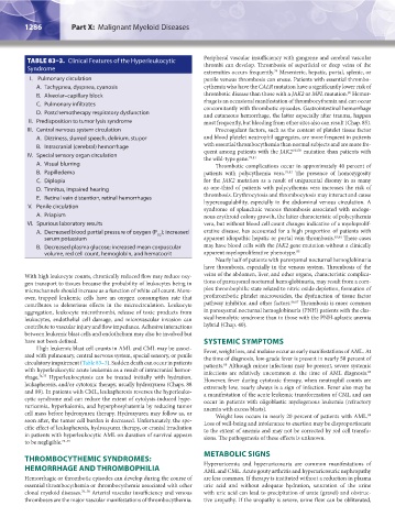
TABLE 83–3. Clinical Features of the Hyperleukocytic Peripheral vascular insufficiency with gangrene and cerebral vascular
thrombi can develop. Thrombosis of superficial or deep veins of the
Syndrome
extremities occurs frequently. Mesenteric, hepatic, portal, splenic, or
79
I. Pulmonary circulation penile venous thrombosis can ensue. Patients with essential thrombo-
A. Tachypnea, dyspnea, cyanosis cythemia who have the CALR mutation have a significantly lower risk of
80
B. Alveolar–capillary block thrombotic disease than those with a JAK2 or MPL mutation. Hemor-
C. Pulmonary infiltrates rhage is an occasional manifestation of thrombocythemia and can occur
concomitantly with thrombotic episodes. Gastrointestinal hemorrhage
D. Postchemotherapy respiratory dysfunction and cutaneous hemorrhage, the latter especially after trauma, happen
II. Predisposition to tumor lysis syndrome most frequently, but bleeding from other sites also can result (Chap. 85).
III. Central nervous system circulation Procoagulant factors, such as the content of platelet tissue factor
A. Dizziness, slurred speech, delirium, stupor and blood platelet neutrophil aggregates, are more frequent in patients
B. Intracranial (cerebral) hemorrhage with essential thrombocythemia than normal subjects and are more fre-
V617F
IV. Special sensory organ circulation quent among patients with the JAK2 mutation than patients with
79,81
the wild-type gene.
A. Visual blurring Thrombotic complications occur in approximately 40 percent of
B. Papilledema patients with polycythemia vera. 79,82 The presence of homozygosity
C. Diplopia for the JAK2 mutation as a result of uniparental disomy in as many
D. Tinnitus, impaired hearing as one-third of patients with polycythemia vera increases the risk of
E. Retinal vein distention, retinal hemorrhages thrombosis. Erythrocytosis and thrombocytosis may interact and cause
hypercoagulability, especially in the abdominal venous circulation. A
V. Penile circulation syndrome of splanchnic venous thrombosis associated with endoge-
A. Priapism nous erythroid colony growth, the latter characteristic of polycythemia
VI. Spurious laboratory results vera, but without blood cell count changes indicative of a myeloprolif-
A. Decreased blood partial pressure of oxygen (P ); increased erative disease, has accounted for a high proportion of patients with
O2
serum potassium apparent idiopathic hepatic or portal vein thrombosis. 83,84 These cases
B. Decreased plasma glucose; increased mean corpuscular may have blood cells with the JAK2 gene mutation without a clinically
volume, red cell count, hemoglobin, and hematocrit apparent myeloproliferative phenotype. 85
Nearly half of patients with paroxysmal nocturnal hemoglobinuria
have thrombosis, especially in the venous system. Thrombosis of the
With high leukocyte counts, chronically reduced flow may reduce oxy- veins of the abdomen, liver, and other organs, characteristic complica-
gen transport to tissues because the probability of leukocytes being in tions of paroxysmal nocturnal hemoglobinuria, may result from a com-
microchannels should increase as a function of white cell count. More- plex thrombophilic state related to nitric oxide depletion, formation of
over, trapped leukemic cells have an oxygen consumption rate that prothrombotic platelet microvesicles, the dysfunction of tissue factor
contributes to deleterious effects in the microcirculation. Leukocyte pathway inhibitor, and other factors. 86,87 Thrombosis is more common
aggregation, leukocyte microthrombi, release of toxic products from in paroxysmal nocturnal hemoglobinuria (PNH) patients with the clas-
leukocytes, endothelial cell damage, and microvascular invasion can sical hemolytic syndrome than in those with the PNH-aplastic anemia
contribute to vascular injury and flow impedance. Adhesive interactions hybrid (Chap. 40).
between leukemic blast cells and endothelium may also be involved but
have not been defined. SYSTEMIC SYMPTOMS
High leukemic blast cell counts in AML and CML may be associ- Fever, weight loss, and malaise occur as early manifestations of AML. At
ated with pulmonary, central nervous system, special sensory, or penile the time of diagnosis, low-grade fever is present in nearly 50 percent of
circulatory impairment (Table 83–3). Sudden death can occur in patients patients. Although minor infections may be present, severe systemic
88
with hyperleukocytic acute leukemia as a result of intracranial hemor- infections are relatively uncommon at the time of AML diagnosis.
89
rhage. 74,75 Hyperleukocytosis can be treated initially with hydration, However, fever during cytotoxic therapy, when neutrophil counts are
leukapheresis, and/or cytotoxic therapy, usually hydroxyurea (Chaps. 88 extremely low, nearly always is a sign of infection. Fever also may be
and 89). In patients with CML, leukapheresis reverses the hyperleuko- a manifestation of the acute leukemic transformation of CML and can
cytic syndrome and can reduce the extent of cytolysis-induced hype- occur in patients with oligoblastic myelogenous leukemia (refractory
ruricemia, hyperkalemia, and hyperphosphatemia by reducing tumor anemia with excess blasts).
cell mass before hydroxyurea therapy. Hydroxyurea may follow as, or Weight loss occurs in nearly 20 percent of patients with AML.
89
soon after, the tumor cell burden is decreased. Unfortunately, the spe- Loss of well-being and intolerance to exertion may be disproportionate
cific effect of leukapheresis, hydroxyurea therapy, or cranial irradiation to the extent of anemia and may not be corrected by red cell transfu-
in patients with hyperleukocytic AML on duration of survival appears sions. The pathogenesis of these effects is unknown.
to be negligible. 73–75
METABOLIC SIGNS
THROMBOCYTHEMIC SYNDROMES:
HEMORRHAGE AND THROMBOPHILIA Hyperuricemia and hyperuricosuria are common manifestations of
AML and CML. Acute gouty arthritis and hyperuricosuric nephropathy
Hemorrhagic or thrombotic episodes can develop during the course of are less common. If therapy is instituted without a reduction in plasma
essential thrombocythemia or thrombocythemia associated with other uric acid and without adequate hydration, saturation of the urine
clonal myeloid diseases. 76–78 Arterial vascular insufficiency and venous with uric acid can lead to precipitation of urate (gravel) and obstruc-
thromboses are the major vascular manifestations of thrombocythemia. tive uropathy. If the uropathy is severe, urine flow can be obliterated,
Kaushansky_chapter 83_p1273-1290.indd 1286 9/21/15 11:14 AM