
Page 136 - Williams Hematology ( PDFDrive )
P. 136
110 Part III: Epochal Hematology Chapter 7: Hematology of the Fetus and Newborn 111
parenteral route may result rarely in neuromuscular complications, capillary fragility. The more serious disorders of periventricular–
293
and an association of intramuscular vitamin K prophylaxis and cancer intraventricular hemorrhage and pulmonary hemorrhage probably
in infancy was suggested but not substantiated. Oral administration, are not caused by coagulation disorders, although such disorders may
300
290
however, appears less reliable and may require repeated doses. The increase the bleeding. Hypoxia seems to affect the clotting status of
301
current recommendation of the American Academy of Pediatrics sug- low-birth-weight infants. Many infants with markedly abnormal
gests that vitamin K , 0.5 to 1.0 mg, be administered intramuscularly at prothrombin times have had hypoxia during delivery or shortly there-
1
294
birth. Even the lower (0.5 mg) parenteral dose may be excessive for after. Cardiovascular collapse seen with episodes of cardiac arrest or
296
preterm (<32 weeks’ gestation) infants, although no toxic effects have with profound shock may cause disseminated intravascular coagulation
295
been reported as a result of very high plasma values. Recent data sug- and generalized bleeding. In many sick premature infants, a combina-
gest that 0.2 mg vitamin K may be appropriate prophylaxis for infants tion of shock, sepsis, liver immaturity, hypoxia, and other factors may
delivered at fewer than 32 weeks’ gestation, but additional oral supple- contribute to the pathogenesis of coagulation abnormalities.
296
mentation is needed when feeding is established. A mixed micellar Arterial and venous thromboses are relatively frequent in new-
vitamin K preparation is particularly well absorbed and may permit borns as compared to other age groups, but greater than 90 percent of
1
prophylaxis with a single oral dose, but the efficacy and safety of oral arterial and greater than 80 percent of venous clots are related to cathe-
297
prophylaxis require further study. ters. Spontaneous thromboses are much less common, and most involve
Table 7–6 shows the values for coagulation factors in healthy 30 the renal veins or, rarely, the pulmonary vasculature. Relative hyperco-
302
to 36 weeks’ gestation premature infants. More prominent decreases agulability in the newborn could result from a difference in the vascular
in factors IX, XI, and XII are noted, which tend to prolong the partial endothelium, activation of the coagulation cascade, diminished coagu-
thromboplastin time. Table 7–6 also shows the values for coagulation lation inhibitor activity, or a defect in fibrinolysis. Inhibitors of coagu-
factors in 28 to 31 weeks’ gestation infants. All of the coagulation factors lation include antithrombin, heparin cofactor II, protein C, and protein
are lower at earlier gestational ages. S. 283,303 The levels of proteins C and S, which are vitamin K dependent,
There are no significant differences in mean prothrombin time as well as antithrombin and heparin cofactor II, are low in the newborn;
determinations between 30 and 36 weeks’ gestation premature and they are in a range associated with thrombotic episodes in adults with
303
full-term infants who have not received vitamin K. Premature infants inherited deficiencies. In addition, the presence of factor V Leiden
298
given vitamin K have a longer mean prothrombin time than do term may occur in as many as 6 percent of newborns. This produces resis-
304
infants similarly treated. In some small infants there is no improve- tance to the action of protein C and may heighten the susceptibility to
ment in prothrombin time or levels of prothrombin, and factors VII thrombosis (Chap. 130). Hyperprothrombinemia caused by the 20210A
305
and X after the intramuscular administration of vitamin K. 286,299 These allele prothrombin gene may affect 1 percent of the population, but
results suggest a greater degree of “immaturity” of the liver in the small the elevated prothrombin level predisposing to thrombosis occurs in
infants. older patients. The combined deficiency of these anticoagulant pro-
306
teins may further intensify the thrombotic risk. However, the precise
Bleeding and Thrombosis role of these inhibitors of coagulation in newborn hypercoagulability
Significant bleeding occurs more often in low-birth-weight infants than is uncertain because a proportionate decrease in vitamin K–dependent
in term newborn infants. Increased capillary fragility is frequently found procoagulant factors (II, VII, IX, X) also is present, and an additional
in premature infants in the first 2 days after birth and is not associated inhibitor, α -macroglobulin, is increased (Chap. 130). Table 7–7 shows
2
with thrombocytopenia. Bleeding under the scalp or in other super- the values for plasma inhibitors of coagulation in premature and term
286
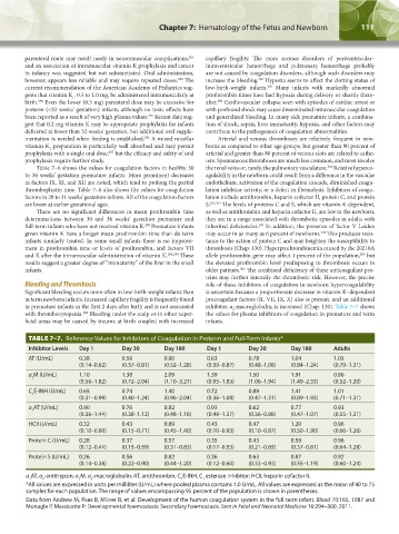
ficial areas may be caused by trauma at birth coupled with increased infants.
TABLE 7–7. Reference Values for Inhibitors of Coagulation in Preterm and Full-Term Infants*
Inhibitor Levels Day 1 Day 30 Day 180 Day 1 Day 30 Day 180 Adults
AT (U/mL) 0.38 0.59 0.90 0.63 0.78 1.04 1.05
(0.14–0.62) (0.37–0.81) (0.52–1.28) (0.39–0.87) (0.48–1.08) (0.84–1.24) (0.79–1.31)
α M (U/mL) 1.10 1.38 2.09 1.39 1.50 1.91 0.86
2
(0.56–1.82) (0.72–2.04) (1.10–3.21) (0.95–1.83) (1.06–1.94) (1.49–2.33) (0.52–1.20)
C E-INH (U/mL) 0.65 0.74 1.40 0.72 0.89 1.41 1.01
1
(0.31–0.99) (0.40–1.24) (0.96–2.04) (0.36–1.08) (0.47–1.31) (0.89–1.93) (0.71–1.31)
α AT (U/mL) 0.90 0.76 0.82 0.93 0.62 0.77 0.93
1
(0.36–1.44) (0.38–1.12) (0.48–1.16) (0.49–1.37) (0.36–0.88) (0.47–1.07) (0.55–1.31)
HCII (U/mL) 0.32 0.43 0.89 0.43 0.47 1.20 0.96
(0.10–0.60) (0.15–0.71) (0.45–1.40) (0.10–0.93) (0.10–0.87) (0.50–1.90) (0.66–1.26)
Protein C (U/mL) 0.28 0.37 0.57 0.35 0.43 0.59 0.96
(0.12–0.44) (0.15–0.59) (0.31–0.83) (0.17–0.53) (0.21–0.65) (0.37–0.81) (0.64–1.28)
Protein S (U/mL) 0.26 0.56 0.82 0.36 0.63 0.87 0.92
(0.14–0.38) (0.22–0.90) (0.44–1.20) (0.12–0.60) (0.33–0.93) (0.55–1.19) (0.60–1.24)
α AT, α -antitrypsin; α M, α -macroglobulin; AT, antithrombin; C E-INH, C esterase inhibitor; HCII, heparin cofactor II.
1 1 2 2 1 1
*All values are expressed in units per milliliter (U/mL) where pooled plasma contains 1.0 U/mL. All values are expressed as the mean of 40 to 75
samples for each population. The range of values encompassing 95 percent of the population is shown in parentheses.
Data from Andrew M, Paes B, Milner B, et al: Development of the human coagulation system in the full-term infant. Blood 70:165, 1987 and
Monagle P, Massicotte P: Developmental haemostasis: Secondary haemostasis. Sem in Fetal and Neonatal Medicine 16:294–300, 2011.
Kaushansky_chapter 07_p0097-0118.indd 111 9/18/15 10:13 PM