
Page 1405 - Williams Hematology ( PDFDrive )
P. 1405
1380 Part X: Malignant Myeloid Diseases Chapter 88: Acute Myelogenous Leukemia 1381
in octogenarians (see Fig. 88–1). The exception to this exponential age- variants of AML (see Chap. 83, Table 83–1 and Fig. 83–3). A cogent
related increase in incidence is APL, which does not change greatly in argument has been made that, for practical purposes, a classification
incidence with age. 188 that initially considers morphologic phenotype and immunophenotype
AML accounts for 15 to 20 percent of the acute leukemias in chil- is advisable. Cytogenetics, molecular genetics, gene-expression profil-
dren and 80 percent of the acute leukemias in adults. It is slightly more ing, and other considerations can, and should, be layered on as available
common in males. Little difference in incidence is seen between indi- and useful in influencing therapy, and these features are starting to be
viduals of African or European descent at any age. A somewhat lower incorporated into the World Health Organization (WHO) Classifica-
189
incidence is seen in persons of Asian descent. An increase in the fre- tion of AML. It is anticipated that molecular classifications will con-
196
quency of AML is seen in Jews, especially those of Eastern European tinue to evolve and dominate clinical decision making in the future. 105
descent. The acute promyelocytic variant of AML is somewhat more
common in Latinos. 190,191 In a large population study of 426,068 patients CLINICAL FEATURES
treated with chemotherapy for malignancy, 301 AML cases occurred,
4.7 times the number expected. Over time (1975 to 2008), the risks SIGNS AND SYMPTOMS
increased for non-Hodgkin lymphoma, declined for ovarian cancer and
myeloma, and were heterogeneous for breast and Hodgkin lymphoma, General
reflecting changing treatment patterns. 192 Signs and symptoms that signal the onset of AML include pallor,
fatigue, weakness, palpitations, and dyspnea on exertion. The signs and
CLASSIFICATION symptoms reflect the development of anemia; however, weakness, loss
of sense of well-being, and fatigue on exertion can be disproportionate
Variants of AML can be identified by morphologic features of blood to the severity of anemia. 197–201
films using polychromatic stains and histochemical reactions, Easy bruising, petechiae, epistaxis, gingival bleeding, conjunctival
193
194
monoclonal antibodies against surface markers, or by the presence hemorrhages, and prolonged bleeding from skin injuries reflect throm-
of specific chromosome translocations or other molecular changes as bocytopenia and are frequent early manifestations of the disease. Very
discussed above. 104,105 The epitopes on the progenitor cells of several infrequently, gastrointestinal, genitourinary, bronchopulmonary, or
phenotypic variants overlap, and several monoclonal antibodies are CNS bleeding occurs at the onset of disease.
required to make specific distinctions among cell types (Table 88–3; see Pustules or other minor pyogenic infections of the skin and of
also “Morphologic Variants of Acute Myelogenous Leukemia” below). minor cuts or wounds are most common. Major infections, such as
Correlation between morphologic and immunologic phenotyping of sinusitis, pneumonia, pyelonephritis, and meningitis, are uncommon
AML is poor. However, poor correlation is expected because morpho- presenting features of the disease, partly because absolute neutrophil
9
logic phenotyping is more subjective, given to observer variation, and counts less than 0.5 × 10 /L are uncommon until chemotherapy starts.
is based on qualitative factors, whereas the immunologic phenotyping, With intensification of neutropenia and monocytopenia after che-
which characterizes surface molecular features, is more accurate and motherapy, major bacterial, fungal, or viral infections become more
reproducible. The correlation is improved only somewhat if morphology frequent. Anorexia and weight loss are frequent findings. Fever is pres-
and histochemistry are coupled. Gene-expression profiling is early in ent in many patients at the time of diagnosis. 200,202–204 Palpable sple-
195
its use as a classification technique for AML but will be more specific nomegaly or hepatomegaly occurs in approximately one-quarter of
and informative than current methods. 104,105 The outcome will depend patients. 197,198,201 Lymphadenopathy is extremely uncommon, 201,205,206
on the simplification and automation of such techniques, and the avail- except in the monocytic variant of AML. 207
ability of drugs that make such distinctions in the prognostic category
of practical utility. Chapter 83 contains the classification of morphologic SPECIFIC ORGAN SYSTEM INVOLVEMENT
Leukemic blast cells circulate and enter most tissues in small numbers.
Occasionally, biopsy (or autopsy) uncovers marked aggregates or infil-
trates of leukemic cells. Collections of such cells may cause functional
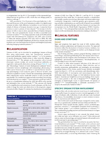
TABLE 88–3. Immunologic Phenotypes of Acute Myelog- disturbances. Extramedullary involvement is most common in mono-
enous Leukemia cytic or myelomonocytic leukemia. 208,209
Phenotype Usually Positive Skin involvement may be of three types: nonspecific lesions, leuke-
mia cutis, or granulocytic (myeloid) sarcoma of skin and subcutis. 210–213
Myeloblastic CD11b, CD13, CD15, CD33, CD117, Nonspecific lesions include macules, papules, vesicles, pyoderma gan-
HLA-DR
grenosum, vasculitis, 214–216 neutrophilic dermatitis (Sweet syndrome),
217
Myelomonocytic CD11b, CD13, CD14, CD15, CD32, CD33, cutis vertices gyrata, and erythema multiforme or nodosum. 211,212
218
HLA-DR Skin involvement preceding marrow and blood involvement or relapse
Erythroid Glycophorin, spectrin, ABH antigens, occurs, but is rare. 219–222
carbonic anhydrase I, HLA-DR, CD71 Sensory organ involvement is very unusual, but retinal, choroi-
(transferrin receptor) dal, iridial, and optic nerve infiltration can occur. Otitis externa and
223
Promyelocytic CD13, CD33 interna, inner ear hemorrhage, and mastoid tumors with seventh nerve
involvement may be presenting signs. 224–226
Monocytic CD11b, 11c, CD13, CD14, CD33, CD65, The gastrointestinal tract may be involved at any point, but func-
HLA-DR
tional disturbances are unusual. 227,228 The mouth, colon, and anal canal
Megakaryoblastic CD34, CD41, CD42, CD61, anti–von are sites of involvement that most commonly lead to symptoms. Oral
Willebrand factor manifestations may prompt the patient to visit the dentist. Gingival or
Basophilic CD11b, CD13, CD33, CD123, CD203c periodontal infiltration and dental abscesses may lead to an extraction,
229
Mast cell CD13, CD33, CD117 followed by prolonged bleeding of an infected tooth socket. Ileoty-
phlitis (enterocolitis), a necrotizing inflammatory lesion involving the
Kaushansky_chapter 88_p1373-1436.indd 1380 9/21/15 11:00 AM