
Page 1439 - Williams Hematology ( PDFDrive )
P. 1439
1414 Part X: Malignant Myeloid Diseases Chapter 88: Acute Myelogenous Leukemia 1415
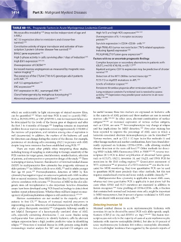
TABLE 88–11. Prognostic Factors in Acute Myelogenous Leukemia (Continued)
Microsatellite instability 1239 (may not be independent of age and High Nrf2 and high ROS expression 1254,1255
t-AML) Overexpression of IL-1 receptor accessory
AC133 expression (shorter remissions and disease-free protein 1256
survival) 1240 Survivin expression in CD34+CD38– cells 1257
Constitutive activity of signal transducer and activator of tran- High TRAIL-R3 (tumor necrosis factor [TNF]-related apoptosis-
scription 3 protein (shorter disease-free survival) 1241 inducing ligand) expression 1258
BAALC gene expression 1242 PICALM-MLLT10 fusion gene expression 302
High S-phase activity in cells surviving after 7 days of induction 1243 Factors with no or uncertain prognostic findings
High EVI1 expression 1244,1245 Complex karyotype or secondary aberrations in patients with
Overexpression of CXCR4 1246 t(8;21), inv(16) t(16;16), or t(9;11) 1259
Increased marrow angiogenesis as measured by magnetic reso- Myeloid antigens: CD11b expression may be predictive of shorter
nance imaging 1247 survival 1260
The presence of the CTLA4 CT60 A/G genotype adult patients Detection of the WT1 (Wilms tumor) transcript 1261
with AML 1248 FLT3-ITD or Asp835 mutations in APL 1262
miR 155 upregulaation 1249 Levels of initiator caspase 1263
ERG expression 1250 Persistent thrombocytopenia after remission induction 1264
EVI1 expression in MLL_rearranged AML 1251 Lung resistance protein: Functional test is needed to assess
Clonal heterogeneity by metaphase karyotyping 1252 activity. 1265 Expression may predict poor outcome in de novo
Abnormal expression of FLI1 protein 1253 AML 1193,1266
which are undetectable by light microscopy of stained marrow films, be useful because these two markers are expressed on leukemic cells
can be quantified. 1267 When real-time PCR is used to quantify PML- in the majority of AML patients and these markers are rare in normal
RAR-α, RUNX1/ETO, or CBF-β/MYH11, risk for treatment failure can marrow cells. 1280,1281 In other cases, aberrant combinations of surface
be determined by the levels of the fusion gene at diagnosis and after antigens 1280,1282 or increased expression of various surface antigens,
the first 3 to 4 months of therapy. 1268 Sampling remains an important such as CD34, are seen. 1283 Immunophenotype may change at relapse
problem because marrow aspiration contains approximately 1/10,000 of and has implications for MRD detection. 1284 Five-color staining has
the marrow cell population, and variation among sites of aspiration is been reported to improve the percentage of AML cases in which a
well documented. In addition, the markers of the leukemic cell used leukemia-associated aberrant immunophenotype can be identified. 1285
for detection can change during the course of the disease. For example, Various markers, such as CLL-1 (C-type lectin-like molecule-1) and
persistence of circulating cells containing t(8;21) in patients with AML other lineage markers and marker combinations, have been found aber-
despite long-term remission has been established using PCR. 1269 rantly expressed on leukemic CD34+CD38–, cells allowing residual
There are many other pitfalls when interpreting these studies, disease detection at the stem cell level. 1286 Other methods for detect-
including timing of sampling in relationship to therapy, sensitivity of the ing MRD include MRI; fluorescence DNA in FISH 1287,1288 ; reverse tran-
PCR reaction for target genes, interlaboratory standardization, selection scriptase (RT)-PCR to detect amplification of abnormal fusion genes,
of patients, and retrospective or prospective design of the study. 1270 There such as t(15;17), t(8;21), inversion 16, and 11q23; and DNA PCR for
is emerging evidence, however, that detection of minimal residual disease mutations in the RAS coding regions. 1267 Quantitative assessment of
(MRD) by multiparameter flow cytometry has prognostic relevance in WT1 expression 1289 or presence of a FLT3 mutation 1290 can also be eval-
older patients, 1271 in children with de novo AML, 1272 and in adults younger uated for MDR monitoring. Real-time quantitative PCR can be used
than age 60 years. 1273 Pretransplantation, detection of MRD by flow to quantitate MDR more precisely than other methods, but this test
cytometry has negative impact on outcome in patients with AML in either requires standardized criteria and is not widely available clinically. 1291
first or second remission. Even MRD levels less than 0.1 percent have an Multiparameter flow cytometry is applicable to most AML cases,
adverse correlation with outcome. 1274 Detection of MRD in the after allo- whereas real-time quantitative PCR is applicable in just above half the
geneic stem cell transplantation is also important. Sensitive chimerism cases when NPM1 and FLT3 mutations are examined in addition to
assays have been developed using PCR-based technology to detect short fusion oncogenes. 1292 Gene profiling of CD34+CD38– cells, a fraction
tandem-repeat polymorphisms. Whether these will have impact on man- that contains both normal and leukemic stem cells, might be important
agement of posttransplantation relapses is still undetermined. 1275,1276 in MRD measurement, but 34 percent of genes modulated in AML stem
Marrow examinations are not needed in the majority of AML cells are shared with normal stem cells. 1293
patients in first CR. 1277 Because of increased myeloid precursors in
regenerating marrow, detection of residual disease may be difficult early Detecting Inversion 16
after a given therapeutic modality. 1278 Cytogenetic followup usually is Minimal residual disease in acute myelomonocytic leukemia with
not helpful. Emergence of a karyotypically unrelated clone of AML inversion 16 can be detected by nested PCR with allele-specific ampli-
cells, especially containing chromosome 7, can occur. Studies using fications (CBF-β on 16q and MYH11 on 16p). 1294,1295 This fusion tran-
multiparameter flow cytometry to identify leukemic cells by aberrant script occurs not only in the majority of cases of acute myelomonocytic
antigen expression have a high positive predictive value in identifying leukemia with marrow eosinophilia (M4Eo), but also in 10 percent of
relapse. 1279 Detection of residual disease in AML patients using double acute myelomonocytic leukemia M4 without eosinophilic abnormali-
immunologic marker analysis for TdT and myeloid CD antigens can ties, a much higher incidence than suggested by the sporadic reports of
Kaushansky_chapter 88_p1373-1436.indd 1414 9/21/15 11:02 AM