
Page 1590 - Williams Hematology ( PDFDrive )
P. 1590
1564 Part XI: Malignant Lymphoid Diseases Chapter 94: Large Granular Lymphocytic Leukemia 1565
HISTOLOGIC AND receptor (KIR) expression. The neoplastic NK cells often demonstrate
37
abnormal KIR expression with either complete absence of surface
IMMUNOPHENOTYPIC FEATURES KIR or restricted KIR expression indicating outgrowth of a clonal
population. 38
The marrow biopsy in T-LGLL may be hypo-, normo-, or hypercellular,
with often preserved trilineage hematopoiesis. There may be occasional
nodules of reactive CD4+ and B lymphocytes as well as scattered LGL,
which are better seen in the aspirate. The presence of interstitial and/or CLINICAL AND LABORATORY
intrasinusoidal clusters of at least eight CD8+ and/or TIA-1+ LGLs or at FEATURES AT PRESENTATION
least six granzyme B+ LGLs has been correlated with marrow involve-
ment by LGLL. Various superimposed findings may reflect secondary Table 94–1 summarizes the clinical features of T-LGLL, as reported
31
immune diseases such as granulocyte maturation arrest and absence of in six large published series. Men and women are equally affected and
red cell precursors (red cell aplasia). T-LGL leukemia invariably affects median age at diagnosis is approximately 60 years. Only a minority of
the spleen, where the major findings are leukemic cell infiltration of patients are less than 50 years old. Approximately one-third of patients
the red pulp cords and sinuses, plasma cell hyperplasia, and prominent with LGLL are asymptomatic at presentation and the diagnosis is made
germinal centers (Fig. 94–1). Hepatic sinusoids and portal areas are on an examination of the blood. The remainder of the patients typically
3,32
infiltrated by LGL. Lymph nodes usually are not involved but can have present with symptoms related to neutropenia (80 percent), anemia
expanded paracortical areas containing plasma cells and LGLs. (45 percent), or both. B symptoms are present only in 15 percent of
T-LGLL and CLPD-NK share overlapping immunophenotypic patients. Physical examination reveals mild to moderate splenomegaly
features in that both often express the NK-associated markers CD16 in 35 percent and hepatomegaly in up to 20 percent. Lymphadenopa-
and CD57. Aberrant expression of NKp46 (CD335), which is normally thy and skin involvement are rare. Pulmonary hypertension develops in
30
selectively expressed by NK cells, occurs in T-LGLL. CD56, which is occasional cases. Rheumatoid arthritis is often a prominent feature of
33
constitutively expressed by circulating NK cells in healthy individuals, LGLL, sometimes resulting in a clinical picture that is difficult to distin-
may be downregulated in CLPD-NK, and its expression in T-LGLL may guish from that of Felty syndrome (FS) (chronic arthritis, splenomegaly,
be associated with a less-favorable clinical course. T-leukemic LGLs and granulocytopenia in the background of longstanding seropositive
34
39
usually are CD3+, CD4–, CD8+, CD16+, CD56–, CD57+, and often rheumatoid arthritis) (Chaps. 56 and 65) . Clonal proliferations of LGL
39
human leukocyte antigen (HLA)-DR+. Less commonly, leukemic LGLs have been observed in patients with FS, and it is likely that a significant
express CD4 with variable CD8 expression. Leukemic T-LGL usually subset of patients diagnosed with FS may in fact have T-LGLL. The dis-
35
express the TCR αβ+ heterodimer, although cases with similar clinical tinction between FS and T-LGLL in patients with rheumatoid arthritis
features have been described that express the γδ TCR heterodimer. In con- has generally been based on whether the LGL proliferation was mono-
36
trast to normal LGL of T-cell origin, leukemic LGL express significantly clonal (LGLL) or polyclonal (FS). However, this distinction cannot
lower levels of CD5 and show abnormal killer immunoglobulin- like always be made, and recent cases of FS with somatic STAT3 mutations
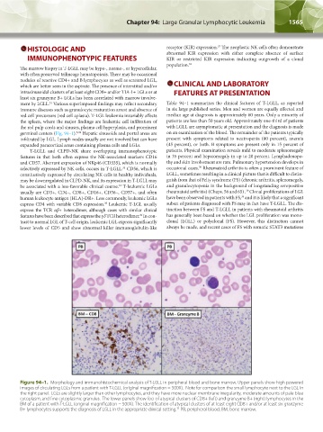
Figure 94–1. Morphology and immunohistochemical analysis of T-LGLL in peripheral blood and bone marrow. Upper panels show high powered
images of circulating LGLs from a patient with T-LGLL (original magnification = 500X). Note for comparison the small lymphocyte next to the LGL in
the right panel. LGLs are slightly larger than other lymphocytes, and they have more nuclear membrane irregularity, moderate amounts of pale blue
cytoplasm, and fine cytoplasmic granules. The lower panels show foci of atypical clusters of CD8+ (left) and granzyme B+ (right) lymphocytes in the
BM of a patient with T-LGLL (original magnification = 500X). The identification of atypical clusters of at least eight CD8+ and/or at least six granzyme
B+ lymphocytes supports the diagnosis of LGLL in the appropriate clinical setting. PB, peripheral blood; BM, bone marrow.
31
Kaushansky_chapter 94_p1563-1568.indd 1565 9/18/15 10:53 AM