
Page 1595 - Williams Hematology ( PDFDrive )
P. 1595
1570 Part XI: Malignant Lymphoid Diseases Chapter 95: General Considerations for Lymphomas 1571
Cancer Institute attempted to reconcile the large number of competing response criteria for NHL were initially published in 1999 by the
6
classifications then in use. The Working Formulation was clinically use- National Cancer Institute Working Group and later revised in 2007
13
ful and gained wide popularity. It divided the specific subtypes among to incorporate positron emission tomography (PET), marrow immu-
14
high-grade, intermediate-grade, and low-grade lymphomas, focusing in nohistochemistry and flow cytometry for response assessment. PET/
part on the expected rate of progression, and not just on the pheno- CT imaging rendered the CRu designation obsolete in 2007, because
type of the case in question. With advances in our understanding of the residual radiographic abnormalities could be accurately determined
immune system and lymphocyte progenitor developmental sequences, to be either residual active lymphoma or posttreatment fibrosis, based
and the availability of monoclonal antibodies for subtyping lymphoid on the metabolic activity of the lesions. These criteria were critically
cells and lymphocyte gene profiling, a new classification schema became reviewed and analyzed by working groups at the 11th and 12th Interna-
possible based on cell type, tissue of origin, immunophenotype, and tional Conferences on Malignant Lymphoma in Lugano, Switzerland,
genotype. in 2011 and 2013 and at the 4th International Workshops on Positron
In 1994, a revised European-American classification of lymphoid Emission Tomography in Lymphoma in Menton, France in 2012. 15,16
neoplasms (the REAL classification) was proposed by the International These international workshops were attended by leading hematolo-
7
Lymphoma Study Group (Chaps. 90 and 96). This group distinguished gists, oncologists, radiation oncologists, pathologists, radiologists, and
three major categories of lymphoid malignancies, which included B-cell, nuclear medicine physicians, representing all major lymphoma clinical
T-cell, and Hodgkin lymphoma. Lymphomas were defined by morpho- trials groups and cancer centers in North America, Europe, Japan, and
logic, immunologic, and genetic techniques. Many of the lymphomas Australasia. Their deliberations culminated in the publication in 2014
were associated with distinct clinical presentations, and cases that did of improved criteria for the initial evaluation, staging and response
not fit into defined entities were left unclassified. Further subclassifi- assessment for both HL and NHL that are relevant for community
17
cation divided each of the B-cell and T-cell lineages into (1) indolent physicians, investigator-led trials, cooperative groups, and registration
8
lymphomas (low risk of rapid progression), (2) aggressive lymphomas trials. These new rules, known as the “Lugano Classification,” depart
(intermediate risk of progression), and (3) very aggressive lympho- substantially from older staging and evaluation systems as detailed
mas (high risk of progression). In 1995, a collaborative project of the later in this chapter.
European Association for Haematopathology and the Society for
Hematopathology began to revise the REAL classification. In 2001, EPIDEMIOLOGY
they published the World Health Organization (WHO) Classification of
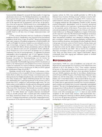
Tumors of the Haematopoietic and Lymphoid Tissues that is used in this Approximately 79,990 new cases of lymphoma were projected to be
chapter and represents the current worldwide consensus classification diagnosed and approximately 20,170 persons in the United States were
18
of malignancies that arise in a lymphocyte. In 2008, the WHO updated expected to die of this disease in 2014. These numbers represent
this classification (Chaps. 90 and 96). 9,10 4.8 percent of the annual incidence of all cancers and 3.4 percent of
Emerging concepts in the staging, therapy, and response evalua- annual cancer-related deaths. The most recent age-adjusted incidence
tion of lymphomas evolved in parallel with the changing nomenclature. rates per 100,000 population provided by Surveillance, Epidemiology,
Initial efforts to define the extent of disease involvement were under- and End Results (SEER) Program of the United States National Cancer
taken primarily for Hodgkin lymphoma (HL) and ultimately led to the Institute are: 24.9 for white males, 17.4 for black males, 17.2 for white
11
Ann Arbor classification, which subdivided patients into four stages, females, and 11.9 for black females. The increased risk for men is sim-
19
each subclassified into A and B groups based on the presence of fevers ilar to that found in other countries, although the incidence of NHL in
greater than 38.3°C, weight loss, and drenching night sweats. The Ann the United States is approximately threefold that of several underdevel-
Arbor system proved useful for decades and was subsequently applied oped countries and twofold that of several comparable industrialized
20
also to non-Hodgkin lymphomas (NHLs). The Cotswold modification countries. The risk of developing lymphoma is less in the United States
12
of the Ann Arbor Classification first formally incorporated computed among persons of African descent in comparison to those of European
tomography (CT) into clinical staging paradigms and introduced the descent. An exponential increase in incidence in NHL among men and
terms “X” for bulky disease and “complete remission unconfirmed” women occurs with increasing age (Fig.95–1). There are some notable
(CRu) to describe patients with a residual mass after treatment that exceptions to this overall trend, however, with lymphoblastic lym-
most likely represented fibrous scar tissue. The sensitivity and accu- phoma occurring most commonly in children, Burkitt lymphoma in the
racy of CT imaging of the abdomen rendered the “staging laparotomy” 20- to 64-year-old age group, and primary mediastinal B-cell lymphoma
for assessment of the abdomen obsolete. The first universally accepted developing at a median age of 35 years. 20
160 Figure 95–1. The graph depicts the rate of increase with age in non-
Hodgkin lymphoma incidence among American males and females.
140 Male This pattern is true for Americans of European or of African descent.
(Data from the Surveillance, Epidemiology, and End Results (SEER) Pro-
120
gram (www.seer.cancer.gov) Research Data (1973-2011), National Can-
Incidence rate (cases/100,000 population) 100 Female cer Institute, DCCPS, Surveillance Research Program, Surveillance Systems
Branch, released April 2014, based on the November 2013 submission.;
80
2014.)
60
40
20
0
< 20 20–49 50–64 65–74 75+
Age interval (years)
Kaushansky_chapter 95_p1569-1586.indd 1570 9/21/15 12:16 PM