
Page 1690 - Williams Hematology ( PDFDrive )
P. 1690
1664 Part XI: Malignant Lymphoid Diseases Chapter 101: Marginal Zone B-cell Lymphomas 1665
Gastric MALT lymphoma presents with nonspecific dyspepsia, a blastic or plasma cell differentiation. Upon immunohistochemistry,
epigastric pain, and nausea. Chronic bleeding may become evident cells are CD20+, CD79a+, CD21+, and CD35+, lacking CD5, CD23,
with progressively worsening iron-deficiency anemia. The antrum is the and CD10, typically expressing immunoglobulin (Ig) M (less often IgA
most frequently involved portion of the organ, although any part of the or IgG), with an immunoglobulin light-chain restriction.
stomach can be affected: intragastric nodularities, enlarged rugal folds,
thickened gastric walls, irregularly shaped superficial erosions or shal- DIFFERENTIAL DIAGNOSIS
low ulcers all represent macroscopic features of this lymphoma.
MALT lymphomas of the ocular adnexa most often arise in the A distinction between reactive (i.e., acquired) and neoplastic MALT
orbit (40 percent of cases), with masses that cause progressive proptosis, is sometimes difficult, especially for MALT lymphoma at early stages
periorbital edema and abnormalities in ocular motility and vision. The of evolution. The demonstration of clonality by virtue of light-chain
conjunctiva is the site of origin in approximately 35 to 40 percent of restriction (on immunohistochemistry or flow cytometry) is of great
the cases, with bilateral involvement observed in nearly 15 percent of diagnostic value. Other entities to be considered are other indolent
patients. More rarely, the lymphoma originates from the lacrimal gland B-cell lymphomas involving extranodal sites, such as mantle cell lym-
(10 percent of the cases) or the eyelid. phoma, small lymphocytic lymphoma and follicular lymphoma. An
Cutaneous MALT lymphoma generally presents with papules, extended immunohistochemical panel, including CD5, CD23, CD10,
plaques or nodules mainly involving the trunk and the upper limbs; the and cyclin D1, along with cytogenetic and molecular analysis is required
occurrence of multiple lesions affecting noncontiguous regions is, how- to rule out other diagnoses.
ever, not infrequent. Lesions may show spontaneous remissions, but
cutaneous relapses are the rule. STAGING PROCEDURES
Immunoproliferative small intestinal disease, which is consid-
ered a special variant of intestinal MALT lymphoma, usually manifests A complete and detailed patient’s history and a full physical exam, along
with a severe and unremitting malabsorption. The lymphoma usually with blood studies evaluating renal and liver function, lactate dehy-
remains confined to the upper intestine and the regional lymph nodes, drogenase (LDH), serum protein electrophoresis and immunofixation,
spreading beyond the abdomen only in advanced stages of the disease and complete serology for HIV, HCV, and hepatitis B virus are consid-
15
and upon transformation into high-grade lymphoma. ered mandatory. Cryoglobulins should be measured in HCV-positive
individuals. Computed tomography of the neck, chest, abdomen, and
pelvis, as well as marrow aspiration and biopsy, are required. H. pylori
MORPHOLOGY status should always be evaluated on the gastric biopsy or through a
The neoplastic lymphocytes tend to infiltrate the marginal zone around urea breath test, and repeated after therapy if positive at baseline. Endo-
reactive B-cell follicles, external to an intact follicular mantle. Their cyto- scopic ultrasound to evaluate the regional lymph nodes and gastric wall
plasm is pale, and they display small to medium-sized and irregularly infiltration is also recommended in gastric MALT lymphoma. Fluores-
shaped nuclei, with dispersed chromatin and inconspicuous nucleoli, cence in situ hybridization for t(11;18) is optional, but useful to guide
either resembling germinal center centrocytes or monocytoid elements. therapy. Involvement with C. jejuni, C. psittaci, or B. burgdorferi may
Scattered, centroblast-like large cells may be present, although never be detected in tumor biopsies from intestine, ocular adnexa or skin,
predominant, as well as mature plasma cells in up to a third of cases. respectively, by polymerase chain reaction, immunohistochemistry or
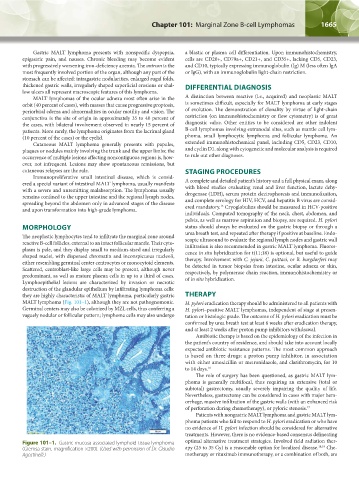
Lymphoepithelial lesions are characterized by invasion or necrotic of in situ hybridization.
destruction of the glandular epithelium by infiltrating lymphoma cells:
they are highly characteristic of MALT lymphoma, particularly gastric THERAPY
MALT lymphoma (Fig. 101–1), although they are not pathognomonic. H. pylori eradication therapy should be administered to all patients with
Germinal centers may also be colonized by MZL cells, thus conferring a H. pylori–positive MALT lymphomas, independent of stage at presen-
vaguely nodular or follicular pattern; lymphoma cells may also undergo tation or histologic grade. The outcome of H. pylori eradication must be
confirmed by urea breath test at least 6 weeks after eradication therapy,
and at least 2 weeks after proton pump inhibitors withdrawal.
Antibiotic therapy is based on the epidemiology of the infection in
the patient’s country of residence, and should take into account locally
expected antibiotic resistance patterns. The most common approach
is based on three drugs: a proton pump inhibitor, in association
with either amoxicillin or metronidazole, and clarithromycin, for 10
to 14 days. 16
The role of surgery has been questioned, as gastric MALT lym-
phoma is generally multifocal, thus requiring an extensive (total or
subtotal) gastrectomy, usually severely impairing the quality of life.
Nevertheless, gastrectomy can be considered in cases with major hem-
orrhage, massive infiltration of the gastric walls (with an enhanced risk
of perforation during chemotherapy), or pyloric stenosis. 17
Patients with nongastric MALT lymphoma and gastric MALT lym-
phoma patients who fail to respond to H. pylori eradication or who have
no evidence of H. pylori infection should be considered for alternative
treatments. However, there is no evidence-based consensus delineating
Figure 101–1. Gastric mucosa-associated lymphoid tissue lymphoma optimal alternative treatment strategies. Involved field radiation ther-
(Giemsa stain, magnification ×200). (Used with permission of Dr. Claudio apy (25 to 35 Gy) is a reasonable option for localized disease. 18,19 Che-
Agostinelli.) motherapy or rituximab immunotherapy, or a combination of both, are
Kaushansky_chapter 101_p1663-1670.indd 1665 9/18/15 9:37 AM