
Page 173 - Williams Hematology ( PDFDrive )
P. 173
148 Part IV: Molecular and Cellular Hematology Chapter 10: Genetic Principles and Molecular Biology 149
complete set of genes (see Fig. 10–1). Occasionally, however, an error AUTOSOMAL DOMINANT INHERITANCE
occurs and pairing during meiosis is imperfect. Under these circum- Diseases caused by autosomal dominant genes are rare, with the most
stances—unequal crossing over (see Fig. 10–5)—one of the daughter common occurring in fewer than 1 in 500 individuals. Therefore, it is
chromosomes contains a duplicated gene, while the other one exists uncommon for two individuals who are both affected by the same auto-
with a gene deleted. Once a duplication has occurred, further dupli- somal dominant disease to produce offspring together. Affected offspring
cations occur more readily, because pairing of the first of the duplicate are usually produced by the union of a normal parent with an affected
genes on one chromosome with the second gene of the duplicate on heterozygous parent. The affected parent can pass either a disease gene
the other produces one chromosome with a triplicated gene and one or a normal gene to the next generation. On average, half the children
with a single gene (Chap. 48). Duplication has probably played a very will be heterozygous and will express the disease, and half will be normal.
important role in the course of evolution because the presence of two The pedigree in Fig. 10–2 shows the transmission of an autoso-
11
genes with the same function allows experiments of nature: Mutations mal dominant trait or disease. Several important characteristics of this
can accumulate on one of the genes while the original function is still pedigree support the conclusion that the trait is inherited in autosomal
provided by the duplicate. Examples of the results of gene duplication dominant fashion:
abound in hematology, particularly with respect to the hemoglobin loci.
The γ-chain loci are duplicated, and there are also two nearly identical 1. The two sexes exhibit the trait in approximately equal proportions,
copies of the α-chain locus (Chap. 48). Furthermore, the close similarity and males and females are equally likely to transmit the trait to their
of their amino acid sequence and the fact that they are tightly linked offspring.
indicate that the β, γ, and δ loci represent the result of duplication of a 2. No generations are skipped. If an individual has the trait, one parent
single ancestral gene. The process of unequal crossing over takes place must also have it. If neither parent has the trait, none of the children
not only between genes, but also within genes. When this occurs, one have it (with the exception of new mutations, as discussed later in
would anticipate that a portion of the amino acid sequence of a protein this section).
is represented twice on one chromosome and is missing on the other. 3. Affected heterozygous individuals transmit the trait to approxi-
The Lepore hemoglobins, leading to a thalassemic clinical state, are an mately half their children, and because gamete transmission is sub-
example of this type of unequal crossing over (Fig. 48–8). These abnor- ject to chance fluctuations, all or none of the children of an affected
mal hemoglobins have the amino acid sequence of the δ chain at the parent may have the trait. When large numbers of matings of this
amino end, and the sequence of the β chain at the carboxyl end. The type are studied, however, the proportion of affected children closely
complement to this kind of abnormality, the “anti-Lepore” hemoglobin, approaches one-half.
also has also been found (Chap. 49). Similarly, a mutation of the glu- The probability that an at-risk individual (e.g., someone with
cocerebrosidase gene causing Gaucher disease has been found to be the a positive family history) will develop a genetic disease is termed the
12
result of a crossover between the active gene and the pseudogene. The recurrence risk. When one parent is affected by an autosomal dominant
two types of haptoglobin represent an ancestral gene and one in which disease (and is a heterozygote) and the other is unaffected, the recur-
a major part of that gene has been duplicated. 13
rence risk for each child is one-half.
An important principle is that each birth is an independent event,
TRANSMISSION OF GENETIC DISEASES much like a coin toss. Thus, even though parents may have already had
a child with the disease, their recurrence risk remains one-half. Even if
The known single-gene diseases can be classified into four major modes they have had several children, all affected (or all unaffected) by the dis-
of inheritance: autosomal dominant, autosomal recessive, X-linked ease, the law of independence dictates that the probability that their next
dominant, and X-linked recessive. The first two types involve genes child will have the disease is still one-half. Parents’ misunderstanding of
14
known to occur on the 22 pairs of autosomes. The last two types occur this principle is a common problem encountered in genetic counseling.
on the X chromosome; very few disease-causing genes are found on the If a child is born with an autosomal dominant disease and there is
Y chromosome. no history of the disease in the family, the child is probably the prod-
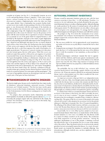
The pedigree chart summarizes family relationships and shows uct of a new (or de novo) mutation. The gene transmitted by one of
16
15
which members of a family are affected by a genetic disease (Fig. 10–2). the parents has thus undergone a mutation from a normal to a disease-
Generally, the pedigree begins with one individual in the family, the causing allele. The genes at this locus in most of the parent’s other germ
proband. This individual is usually the first person in the family diag- cells are still normal. In this situation the recurrence risk for the parent’s
nosed or seen in a clinic. subsequent offspring is not greater than that of the general population.
The offspring of the affected child, however, will have a recurrence risk
of one-half. Because these diseases often reduce the potential for repro-
duction, many autosomal dominant diseases result from new mutations.
Occasionally, two or more offspring have symptoms of an auto-
Aa aa somal dominant disease when there is no family history of the disease.
Because mutation is a rare event, it is unlikely that this disease would
be a result of multiple mutations in the same family. The mechanism
17
aa aa Aa aa most likely responsible is termed germline mosaicism. During the
embryonic development of one of the parents, a mutation occurred
that affected all or part of the germline, but few or none of the somatic
cells of the embryo. Thus, the parent carries the mutation in the parent’s
aa aa aa Aa aa Aa
germline but does not actually express the disease. As a result, the unaf-
Figure 10–2. Pedigree for an autosomal dominant disease. (Repro- fected parent can transmit the mutation to multiple offspring. This
duced with permission from Jorde LB, Carey JC, Barnshad MJ: Medical phenomenon, although relatively rare, can have significant effects on
Genetics, 4th edition. Philadelphia, PA: Mosby/Elsevier, 2010.) recurrence risks. 18
Kaushansky_chapter 10_p0143-0154.indd 148 9/18/15 10:22 PM