
Page 174 - Williams Hematology ( PDFDrive )
P. 174
148 Part IV: Molecular and Cellular Hematology Chapter 10: Genetic Principles and Molecular Biology 149
AUTOSOMAL RECESSIVE INHERITANCE If two parents both have a recessive disease, they each must be
Like autosomal dominant diseases, diseases caused by autosomal reces- homozygous for the disease. Therefore, all their children also must be
sive genes are rare in populations, although there can be numerous car- affected. This distinguishes recessive from dominant inheritance because
riers. Sickle cell disease is seen in approximately 1 in 600 Americans of two parents both affected by a dominant gene are nearly always both
African descent, but it occurs in the heterozygote state in approximately heterozygotes and thus one-fourth of their children will be unaffected.
1 in 12 members of this population. Under most circumstances, car- Because carrier parents usually are unaware that they both carry
19
riers are phenotypically normal. Like autosomal dominant diseases, the same recessive allele, they often produce an affected child before
many autosomal recessive diseases are characterized by incomplete knowing of their condition. Carrier detection tests can identify hete-
penetrance and variable expressivity. rozygotes by measuring the reduced amount of a critical enzyme. This
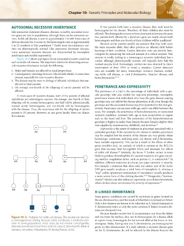
Figure 10–3 shows a pedigree for an autosomal recessive condition enzyme is totally lacking in a homozygous recessive individual, but a
such as sickle cell disease. The important criteria for discerning autoso- carrier, although phenotypically normal, will typically have half the
mal recessive inheritance include the following: normal enzyme level. Increasingly, carriers are now detected by direct
examination of their DNA to reveal a mutation. Carrier detection
1. Males and females are affected in equal proportions. tests are available for many hematologic recessive diseases, includ-
2. Consanguinity (marriage between related individuals) is sometimes ing sickle cell disease, α- and β-thalassemia, Gaucher disease, and
present, especially for rare recessive diseases. hemochromatosis. 20–22
3. The disease may be seen in siblings of affected individuals, but usu-
ally not in their parents.
4. On average, one-fourth of the offspring of carrier parents will be PENETRANCE AND EXPRESSIVITY
affected. The penetrance of a trait is the percentage of individuals with a spe-
In most cases of recessive disease, both of the parents of affected cific genotype who also exhibit the expected phenotype. Incomplete
individuals are heterozygous carriers. On average, one-fourth of their penetrance means that individuals who have the gene disease-causing
offspring will be normal homozygotes, one-half will be phenotypically genotype may not exhibit the disease phenotype at all, even though the
normal carrier heterozygotes, and one-fourth will be homozygotes genotype and the associated disease may be transmitted to the next gen-
with the disease. Thus, the recurrence risk for the offspring of carrier eration. Penetrance can increase with age, and it can differ between the
parents is 25 percent. However, in any given family, there are chance sexes. For example, the penetrance of hemochromatosis, an autosomal
fluctuations. recessive condition, increases with age as iron accumulates in organs
such as the heart and liver. The penetrance of the hemochromatosis
genotype is higher in males than females because females deplete their
iron supplies by menstruation, childbirth, and lactation. 23
Expressivity is the extent of variation in phenotype associated with a
Aa AA particular genotype. If the expressivity of a disease is variable, penetrance
may be complete but the severity of the disease can vary greatly. Many
hematologic conditions, including sickle cell disease and β-thalassemia,
have variable expressivity. This can be a result of the effects of other
genes (modifier loci), an example of which is variants in the BCL11A
gene that increase fetal hemoglobin levels and attenuate the effects
of sickle cell disease. Similarly, the factor V Leiden variant is more
24
likely to produce thrombophilia if a second mutation of a gene encod-
ing another coagulation factor, such as protein C, is coinherited. In
25
AA Aa AA Aa AA addition, different mutations at a locus can cause variation in severity.
For example, a mutation that alters only one amino acid of the factor
VIII gene usually produces a mild form of hemophilia A, whereas a
“stop” codon (premature termination of translation) usually produces
a more-severe form of this clotting disorder. 26,27 Nongenetic (“environ-
mental”) factors can also influence expression, as in hemochromatosis,
where alcohol abuse can increase the severity of expression. 28
AA Aa Aa Aa AA
X-LINKED INHERITANCE
Some genetic conditions are caused by mutations in genes located on
the sex chromosomes, and that mode of inheritance is termed sex linked.
Only a few diseases are known to be inherited as X-linked dominant or
Y chromosome traits, so only the more common X-linked recessive dis-
eases are discussed here.
aa Aa aa AA
Because females receive two X chromosomes, one from the father
Figure 10–3. Pedigree for sickle cell disease. The double bar denotes and one from the mother, they can be homozygous for a disease allele
a consanguineous mating. Because sickle cell disease is relatively com- at a given locus, homozygous for the normal allele at the locus, or het-
mon in some populations, most cases do not involve consanguinity. erozygous. Males, having only one X chromosome, are hemizygous for
(Reproduced with permission from Jorde LB, Carey JC, Barnshad MJ: Medical genes on this chromosome. If a male inherits a recessive disease gene
Genetics, 4th edition. Philadelphia, PA: Mosby/Elsevier, 2010.) on the X chromosome, he will be affected by the disease because the
Kaushansky_chapter 10_p0143-0154.indd 149 9/18/15 10:22 PM