
Page 175 - Williams Hematology ( PDFDrive )
P. 175
150 Part IV: Molecular and Cellular Hematology Chapter 10: Genetic Principles and Molecular Biology 151
Y chromosome does not carry a normal allele to counteract the effects Persons with abnormal numbers of X chromosomes, such as those
of the disease gene. Because a single copy of an X-linked recessive gene with Turner syndrome or Klinefelter syndrome, are not physically nor-
will cause disease in a male, whereas two copies are required for disease mal. This situation presents a puzzle because they presumably have only
expression in females, more males are affected by X-linked recessive dis- one active X chromosome, just as individuals with normal numbers of
eases than are females. chromosomes do. This is probably because the distal tips of the short
and long arms of the X chromosome, as well as several other regions on
X INACTIVATION the chromosome arm, are not inactivated. Thus, X inactivation is also
In the late 1950s, Mary Lyon proposed that one X chromosome in the known to be incomplete.
somatic cells of females is permanently inactivated, a process termed Although the mechanisms underlying X inactivation are only
X inactivation (Fig. 10–4). 29–33 This proposal, the Lyon hypothesis, explains partially understood, the gene responsible for initiating X inactivation,
34
why most gene products coded by the X chromosome are present in XIST, has been identified. This gene encodes a long noncoding RNA
equal amounts in males and females, even though males have only one (lncRNA) that coats one of the X chromosomes, which is then inacti-
X chromosome and females have two X chromosomes. This phenome- vated (it is estimated that the human genome contains approximately
1
non is called dosage compensation. The inactivated X chromosomes are 9000 lncRNA genes ). Methylation of X chromosome DNA, a process in
observable in many interphase cells as highly condensed intranuclear which DNA is inactivated when cytosine bases are enzymatically con-
chromatin bodies, termed Barr bodies (after Barr and Bertram, who dis- verted to 5-methylcytosine, occurs on the inactivated X chromosome.
covered them in the late 1940s). Normal females have one Barr body in Inactive X chromosomes can be at least partially reactivated in vitro by
each somatic cell, whereas normal males have no Barr bodies. administering 5-azacytidine, a demethylating agent.
X-inactivation occurs very early in embryonic development—
approximately 7 to 14 days after fertilization. In each somatic cell, one Characteristics of Pedigrees
of the two X chromosomes is inactivated. In some cells, the inactivated X-linked pedigrees show distinctive modes of inheritance. The most
X chromosome is the one contributed by the father; in other cells it is striking characteristic is that females seldom are affected. To express an
the one contributed by the mother. Once the X chromosome has been X-linked recessive trait, a female must be homozygous: either both her
inactivated in a cell, all the descendants of that cell have the same chro- parents are affected, or her father is affected and her mother is a carrier.
mosome inactivated. Thus inactivation is said to be random but fixed. Such matings are rare.
Some individuals do not have the normal number of X chromo- The following are important principles of X-linked recessive
somes in their somatic cells. For example, males with Klinefelter syn- inheritance:
drome typically have two X chromosomes and one Y chromosome. 1. The trait is seen much more often in males than in females.
These males do have one Barr body in each cell. Females whose cell 2. Because a father can give a son only a Y chromosome, the trait is
nuclei have three X chromosomes have two Barr bodies in each cell, never transmitted from father to son.
and females whose cell nuclei have four X chromosomes have three 3. The gene can be transmitted through a series of carrier females, caus-
Barr bodies in each cell. Females with Turner syndrome have only one ing the appearance of one or more “skipped generations.”
X chromosome and no Barr bodies. Thus, the number of Barr bodies is 4. The gene is passed from an affected father to all his daughters, who,
always one less than the number of X chromosomes in the cell. All but as phenotypically normal carriers, transmit it to approximately half
one X chromosome are always inactivated. their sons, who are affected. Good examples of X-linked hematologic
disorders include hemophilia A (clotting factor VIII deficiency),
hemophilia B (clotting factor IX deficiency), and G6PD deficiency.
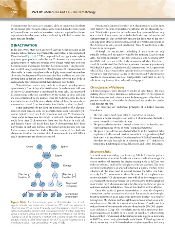
Ovum X m X p Sperm
Recurrence Risks
m p
X X Zygote The most common mating type involving X-linked recessive genes is
the combination of a carrier female and a normal male. On average, the
carrier mother will transmit the disease-causing allele to half her sons
m p
m p
X X X X Embryo (who are affected) and half her daughters (who are carriers). The other
common mating type is an affected father and a normal mother. In this
situation, all the sons must be normal because the father can trans-
Barr body X m X p Barr body mit only his Y chromosome to them. Because all the daughters must
inactive X p inactive X m receive the father’s X chromosome, they will all be heterozygous carri-
ers. Because the sons must receive the Y chromosome and the daughters
X m X m X p X p Polyclonal must receive the X chromosome with the disease gene, these are precise
mosaicism
outcomes and not probabilities. None of the children will be affected.
Once the mode of genetic transmission is clear, the diagnostic
X m X m X m X m X m X m Monoclonal alternatives can be narrowed considerably. For example, methemoglo-
proliferation binemia transmitted as an autosomal dominant disorder is a result of
hemoglobin M, whereas methemoglobinemia transmitted as an auto-
Figure 10–4. The X inactivation process. At fertilization, the female somal recessive disorder is a result of cytochrome b5 reductase (the
zygote inherits one maternal chromosome (X ) and one paternal X reduced form of nicotinamide adenine dinucleotide [NADH] diapho-
m
chromosome (X ). At some time early in embryogenesis, one X in each rase) deficiency (Chap. 49). Hemolytic anemia with autosomal dom-
P
cell is inactivated at random and condenses to form the Barr body. The
active X remains active not only for the lifetime of that cell but for the inant transmission is likely to be a result of hereditary spherocytosis,
lifetime of all of its progeny. A tumor with a clonal origin will consist but sex-linked transmission of the hemolytic state suggests a deficiency
entirely of cells in all of which either X or X is active. A tumor with a of G6PD or, more rarely, phosphoglycerate kinase. A bleeding disorder
P
m
P
m
multicentric origin may contain both X and X cells. that is transmitted in an X-linked fashion may be caused by a deficiency
Kaushansky_chapter 10_p0143-0154.indd 150 9/18/15 10:22 PM