
Page 171 - Williams Hematology ( PDFDrive )
P. 171
146 Part IV: Molecular and Cellular Hematology Chapter 10: Genetic Principles and Molecular Biology 147
5’ 3’ Because there are 64 (4 × 4 × 4) possible codons but only 20 amino
acids, there are many cases in which several codons correspond to the
same amino acid.
The genetic code is universal: All living organisms use precisely the
same DNA codes to specify proteins except for mitochondria, the cyto-
plasmic organelles in which cellular respiration takes place—they have
their own extranuclear DNA. Several codons of mitochondrial DNA
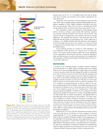
Sugar-phosphate
backbone encode different amino acids than do the same nuclear DNA codons.
DNA replication consists of breaking the weak hydrogen bonds
One helical turn = 34 nm Bases The consistent pairing of adenine with thymine and of guanine with
between the bases, leaving a single strand with each base unpaired.
cytosine, known as complementary base pairing, is the key to accurate
replication. The unpaired base attracts a free nucleotide only if the
nucleotide has the proper complementary base. When replication is
complete, a new double-stranded molecule identical to the original is
formed. The single strand is said to be a template, or molecule on which
a complementary molecule is built, and is the basis for synthesizing the
new double strand.
Several different proteins are involved in DNA replication. The
most important of these proteins is an enzyme known as DNA poly-
merase. This enzyme travels along the single DNA strand, adding the
correct nucleotides to the free end of the new strand and checking to
make sure that its base is actually complementary to the template base.
This mechanism of DNA proofreading substantially enhances the accu-
racy of DNA replication.
MUTATIONS
A mutation is any inherited alteration of genetic material. Mutations
may cause disease or be subtle, silent substitutions that do not change
amino acids. A base pair change that alters a single amino acid is termed
a missense mutation, and a base pair change that produces a premature
stop codon is termed a nonsense mutation. Because they typically result
in a complete loss of gene product, nonsense mutations usually pro-
duce a more-severe disease phenotype than do missense mutations. For
example, nonsense mutations in the factor VIII gene are much more
likely to produce severe hemophilia A than are missense mutations. This
is an example of a genotype-phenotype correlation, which can be use-
ful in predicting the severity of disease. A frameshift mutation involves
the insertion or deletion of one or more base pairs of the DNA mole-
3’ 5’ cule. These mutations change the entire “reading frame” of the DNA
sequence because the deletion or insertion is not a multiple of three base
pairs (bp; the number of base pairs in a codon). Frameshift mutations
Adenine can thus greatly alter the amino acid sequence and typically lead to a
Thymine premature stop codon downstream of the mutation. (“In-frame” inser-
Guanine tions or deletions, in which a multiple of three bases is inserted or lost,
tend to have less-severe disease consequences than do frameshift muta-
Cytosine
tions.) Splice-site mutations describe alterations of the DNA sequence
at intron–exon boundaries (see section on Genes to Proteins above).
Figure 10–1. Watson-Crick model of the DNA molecule. The DNA These result in a mature mRNA transcript that contains introns or lacks
structure illustrated here is based on that published by James Watson exons. Table 10–1 gives examples of hematologic diseases caused by dif-
and Francis Crick in 1953. Note that each side of the DNA molecule con- ferent types of mutations.
sists of alternating sugar and phosphate groups. Each sugar group is
united to the sugar group opposite it by a pair of nitrogenous bases Mutations are rare events. The rate of spontaneous mutations (those
(adenine-thymine or cytosine-guanine). The sequence of these pairs occurring in the absence of exposure to known mutagens) in humans
−4
−6
constitutes a genetic code that determines the structure and func- is approximately 10 to 10 per gene per generation. This rate varies
tion of a cell. (Reproduced with permission from Patton KT, Thibodeau GA: from one gene to another, with larger mutation rates for larger genes. At
Anatomy & physiology, 8th edition. St. Louis, MO:Mosby/Elsevier, 2013.) the nucleotide level, the human mutation rate is approximately 10 per
−8
nucleotide per generation. Certain DNA sequences have particularly
4,5
high mutation rates and are known as mutational hot spots. In particu-
lar, sequences consisting of a cytosine base followed by a guanine base
(CpG) are highly susceptible to mutation and are known to account for
a disproportionately large percentage of disease-causing mutations.
Kaushansky_chapter 10_p0143-0154.indd 146 9/18/15 10:22 PM