
Page 1751 - Williams Hematology ( PDFDrive )
P. 1751
1726 Part XI: Malignant Lymphoid Diseases Chapter 106: Essential Monoclonal Gammopathy 1727
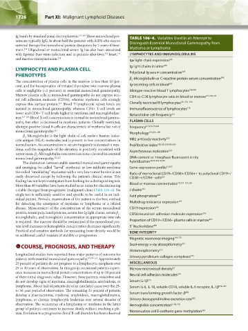
Ig bands by standard zonal electrophoresis. 167,168 These monoclonal pro- TABLE 106–4. Variables Used in an Attempt to
teins are typically IgG. In about half the patients with AIDS who receive
antiviral therapy the monoclonal protein disappears by 5 years of treat- Distinguish Essential Monoclonal Gammopathy from
ment. Oligoclonal or monoclonal serum Ig has also been associated Myeloma or Lymphoma
212
214
213
with Epstein-Barr virus infection and in patients after liver, heart, LYMPHOCYTES AND IMMUNOGLOBULINS
and marrow transplantation. 215 Igκ light-chain expression 245
Ig light chains in urine 245
LYMPHOCYTE AND PLASMA CELL 245
PHENOTYPES Polyclonal Ig serum concentration 246
2
The concentration of plasma cells in the marrow is less than 10 per- β -Microglobulin or C-reactive protein serum concentration
cent, and the incorporation of tritiated thymidine into marrow plasma Ig-secreting cells in blood 247
cells is negligible (<1 percent) in essential monoclonal gammopathy. Idiotype-reactive blood T lymphocytes 248,249
Marrow plasma cells in monoclonal gammopathy do not express neu- CD4-to-CD8 lymphocyte ratio in blood or marrow 217,250,251
ral cell adhesion molecule (CD56), whereas myeloma cells strongly
express this surface protein. Blood T-lymphocyte subset levels are Clonally restricted B lymphocytes 221,252–254
216
normal in monoclonal gammopathy, whereas CD4+ T-cell levels are Immunofluorescence of lymphocytes 218
lower and CD8+ T-cell levels higher in myeloma and macroglobuline- Natural killer cell frequency 255
mia. 217–220 Blood B-cell concentration is normal in monoclonal gammo-
pathy, but often is decreased in myeloma patients. Clonally restricted, PLASMA CELLS
idiotype-positive blood B cells are characteristic of myeloma but not of Frequency 6,7,9,10,15,203
monoclonal gammopathy. 221 Morphology 219,256–258
β -Microglobulin is the light chain of cell surface human leuko-
2
cyte antigen (HLA) molecules and is present in low concentrations in MB2 antibody reactivity 259
normal serum. Its concentration in serum frequently is elevated in mye- Proliferative index 219,220,250,256,260
loma, and the magnitude of the elevation is positively correlated with Asynchronous replication 261
tumor mass. β -Microglobulin concentration is not elevated in essential
2
monoclonal gammopathy. 222,223 DNA content or interphase fluorescent in situ
The distinction between stable essential monoclonal gammopathy hybridization 38,39,43,219,256
and emerging (so-called “larval” myeloma) or low-infiltrate myeloma Gene-expression profile 51,237
(so-called “smoldering” myeloma) with a very low tumor burden is not Ratio of monoclonal CD19–/CD38+/CD56++ to polyclonal CD19+/
easily discerned except by following the patient’s clinical status. This CD38++/CD56– cells 262
finding has not kept investigators from looking for a distinguishing test. 10,219–221,253
More than 40 variables have been studied as an index for discriminating Blood or marrow concentration
a stable (benign) from progressive (malignant) clone (Table 106–4). No J chains 263
single test is sufficiently sensitive and specific to be useful in an indi- Acid phosphatase 264
vidual patient. Periodic examination of the patient is the best method
for detecting the emergence of myeloma or lymphoma or a related Multidrug resistance expression 265
disease. Measurement of the concentration of the serum monoclonal CD19 expression 266
protein, serum polyclonal proteins, serum free Ig light chains, serum β - CD56/neural cell adhesion molecule expression 216
2
microglobulin, and hemoglobin concentration at appropriate intervals
is required. The marrow should be reexamined if the monoclonal pro- Proportion of CD19+/CD56– plasma cells in marrow 267
tein level increases or hemoglobin concentration decreases significantly. 5′ Nucleotidase 268
Practical and sensitive methods for measuring bone density would be BONE INTEGRITY
an additional useful measure of stability or progression.
Magnetic resonance imaging 269,270
COURSE, PROGNOSIS, AND THERAPY Dual-energy x-ray absorptiometry 271
Histomorphometry 272
Longitudinal studies have reported three major patterns of outcome for 273
patients with essential monoclonal gammopathy. 10,224–226 Approximately Urinary pyridinium-collagen complexes
25 percent of patients do not progress to a lymphocytic neoplasm over MISCELLANEOUS
25 to 30 years of observation. In this group, occasional patients experi- Marrow microvessel density 62
ence increases in monoclonal protein concentration of up to 50 percent 274
of their initial diagnostic value. However, these patients restabilize and Neural cell adhesion molecules
do not develop signs of myeloma, macroglobulinemia, amyloidosis, or Serum IL-1β 275
lymphoma. About half of patients die of an unrelated cause over the 25- Serum IL-6, IL-10, soluble CD16, soluble IL-6 receptor, IL-1β 276–281
to 30-year period of observation. The remaining 25 percent of patients 282
develop a plasmacytoma, myeloma, amyloidosis, macroglobulinemia, Serum transforming growth factor-β
lymphoma, or chronic lymphocytic leukemia over several decades of Urinary deoxypyridinoline excretion rate 282
observation. The occurrence of a lymphoma or myeloma in the latter Hemoglobin concentration 8,148,178
group of patients continues to increase slowly without reaching a pla- 283
teau. Evolution to a progressive clonal B-cell disorder has been observed Mononuclear cell E-cadherin gene methylation
Kaushansky_chapter 106_p1721-1732.indd 1726 9/21/15 12:40 PM