
Page 1747 - Williams Hematology ( PDFDrive )
P. 1747
1722 Part XI: Malignant Lymphoid Diseases Chapter 106: Essential Monoclonal Gammopathy 1723
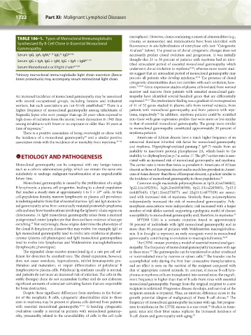
TABLE 106–1. Types of Monoclonal Immunoglobulin (metaphase). However, clones containing numerical abnormalities (e.g.,
trisomy or monosomy) and translocations have been identified with
Synthesized By B-Cell Clone in Essential Monoclonal fluorescence in situ hybridization of interphase cells (see “Cytogenetic
Gammopathy Analysis” below). The presence of clonal cytogenetic changes does not
Serum IgG, IgA, IgM, 6–12 IgE, IgD 64–66 necessarily predict clonal evolution and progression. It was initially
63
Serum IgG + IgA, IgG + IgM, IgG + IgA + IgM 67–70 thought that 25 to 30 percent of patients with myeloma had an iden-
tified antecedent period of essential monoclonal gammopathy which
Serum Monoclonal κ or λ light chain* 71,97 underwent clonal evolution to myeloma 43,46 ; whereas more recent stud-
*Urinary monoclonal immunoglobulin light chain excretion (Bence ies suggest that an antecedent period of monoclonal gammopathy may
Jones proteinuria) may accompany serum monoclonal light chain. precede all patients who develop myeloma. 48,49 The presence of clonal
cytogenetic abnormalities does not correlate with such evolution, how-
ever. 37,38,45 Gene-expression studies of plasma cells isolated from normal
marrow and marrow from patients with essential monoclonal gam-
An increased incidence of monoclonal gammopathy may be associated mopathy have identified several hundred genes that are differentially
with several occupational groups, including farmers and industrial expressed. 50,51 The predominate finding was a gradient of overexpression
workers, but such associations are not firmly established. There is a of 41 of 52 genes studied in plasma cells from normal subjects, from
32
higher frequency of monoclonal gammopathy among inhabitants of patients with monoclonal gammopathy, and from patients with mye-
Nagasaki, Japan who were younger than age 20 years when exposed to loma, respectively. In addition, myeloma patients could be stratified
51
high doses of radiation from the atomic bomb detonation in 1945 than into those with gene-expression profiles that were more or less similar
among inhabitants with lower or no exposure or older than 20 years at to that of essential monoclonal gammopathy. The group more similar
time of exposure. 21 to monoclonal gammopathy constituted approximately 30 percent of
There is a positive association of being overweight or obese with myeloma patients.
the incidence of a monoclonal gammopathy and a similar positive Americans of African descent have a much higher frequency of an
33
association exists with the incidence of or mortality from myeloma. 34–36 autosomal dominant inherited risk factor for monoclonal gammopathy
and myeloma. Hyperphosphorylated paratarg-7 (pP-7) results from an
inability to inactivate protein phosphatase 2A, which leads to the
ETIOLOGY AND PATHOGENESIS inability to dephosphorylate p-7 at serine 17. The pP-7 carrier state is asso-
ciated with an increased risk of monoclonal gammopathy and myeloma.
Monoclonal gammopathy can be compared with any benign tumor, The carrier state is more than twice as prevalent in Americans of African
such as a colonic adenomatous polyp, which can remain the same size descent as those of European descent and is much less prevalent in Ameri-
indefinitely or undergo malignant transformation at an unpredictable cans of Asian descent than those of European descent, a gradient similar to
future time. the incidence of monoclonal gammopathy in those populations. 52
Monoclonal gammopathy is caused by the proliferation of a single Common single nucleotide polymorphisms at 2p23.3(rs6746082),
B lymphocyte, a plasma cell progenitor, leading to a clonal population 3p22.1(rs1052501), 3q26.2(rs10936599), 6p21.33(rs2285803), 7p15.3
that reaches a steady-state at approximately 1 to 5 × 10 cells. At this (rs4487645), 17p11.2(rs4273077), and 22q13.1(rs877529) are associ-
10
cell-population density, marrow lymphocyte or plasma cell prevalence ated with increased risk of myeloma. Similarly these polymorphisms
is indistinguishable from that of normal marrow. IgG and IgA monoclo- independently increased the risk of monoclonal gammopathy. Poly-
nal gammopathy arise from somatically mutated postswitch preplasma morphism associations were independent; risk increased with a larger
cells and may have translocations involving the Ig heavy-chain region on number of risk alleles carried, supporting a polygenic model of disease
chromosome 14. IgM monoclonal gammopathy arises from a mutated susceptibility to monoclonal gammopathy and, therefore, to myeloma. 53
postgerminal center lymphocyte that does not have evidence of isotype MYD88 L265 is a somatic mutation found in approximately
switching. Not surprisingly, these origins determine the phenotype of 50 percent of individuals with IgM monoclonal gammopathy and in
37
the clonal B-lymphocytic diseases that may evolve. For example, IgG or more than 90 percent of patients with Waldenström macroglobuline-
IgA monoclonal gammopathy tend to evolve into myeloma or plasma- mia. It is thought to represent an early oncogenic event in monoclonal
cytoma (plasma cell phenotypes) and IgM monoclonal gammopathies gammopathy contributing to evolution to macroglobulinemia. 54,55
tend to evolve into lymphomas and Waldenström macroglobulinemia The C57BL mouse provides a model of essential monoclonal gam-
(lymphocytic phenotypes). mopathy. The frequency of monoclonal gammopathy increases with age
The expanded clone secretes monoclonal Ig at a rate per cell suf- in these mice. The gammopathy can be transferred to either irradiated
56
ficient for detection by standard tests. The clonal expansion, however, or nonirradiated mice by marrow or spleen cells. The transfer can be
57
does not cause osteolysis, hypercalcemia, inhibit hematopoietic pro- accomplished only during the first four consecutive transplantations,
liferation and maturation, or impair differentiation of polyclonal B and no effect is seen on the survival of the recipient compared with
lymphocytes to plasma cells. Polyclonal Ig synthesis usually is normal, that of appropriate control animals. In contrast, if mouse B-cell lym-
and patients do not incur an increased risk of infection. The cells in the phoma or myeloma cells are transplanted into normal mice, the engraft-
stable (benign) clone do not accumulate further and do not elaborate ment frequency is higher than that of B cells from mice with essential
significant amounts of osteoclast-activating factors that are responsible monoclonal gammopathy. Passage from the original recipient to a new
for bone destruction. recipient is unlimited. Progressive disease develops, and survival of the
Despite these significant differences from myeloma in the behav- recipient animals is impaired. Thus, an intrinsic difference exists in the
ior of the neoplastic B cells, cytogenetic abnormalities akin to those growth potential (degree of malignancy) of these B-cell clones. The
43
seen in myeloma may be present in plasma cells derived from patients frequency of monoclonal gammopathy increases with age, but progres-
with essential monoclonal gammopathy. 37–47 G-banding cytogenetic sion to myeloma in the C57BL mouse is a rare event. Studies in trans-
58
evaluation usually is normal in patients with monoclonal gammop- genic mice and their litter mates replicate the increased incidence of
athy, presumably related to the unavailability of cells in the cell cycle B-cell clones and gammopathy with aging. 59
Kaushansky_chapter 106_p1721-1732.indd 1722 9/21/15 12:39 PM