
Page 1759 - Williams Hematology ( PDFDrive )
P. 1759
1734 Part XI: Malignant Lymphoid Diseases Chapter 107: Myeloma 1735
28
EPIDEMIOLOGY or myeloma after 15 to 20 years, and accelerates MG transformation
29
to myeloma. People exposed to fresh wood, wood dust, or working in
INCIDENCE AND PREVALENCE saw mill factories had an increased risk of myeloma in some studies. 30,31
Myeloma represents the second most common hematologic cancer, Finally, associations between myeloma risk and autoimmune diseases
32,33
or pernicious anemia ) or infections
(especially rheumatoid arthritis
34
accounting for 1.4 percent of all cancers and 10 percent of hemato- (HIV and hepatitis C 35,36 ) have been proposed, based on a retrospec-
logic malignancies, with a prevalence of 83,367 people in 2011. The tive study among American male military veterans, 37,38 suggesting an
2
American Cancer Society has estimated that 24,050 new myeloma cases immune-mediated mechanism for malignant transformation. Human
were diagnosed in the United States in 2014, of which approximately herpes virus 8 DNA sequences responsible for Kaposi sarcoma and Cas-
11,090 will die. Most of the patients are diagnosed among people ages tleman disease pathogenesis have been reported by some investigators
39
65 to 74 years, with a median age at onset of 69 years; only 4 percent of in marrow dendritic cells of myeloma patients, although other studies
cases occur before age 45 years. Men are affected more frequently than suggest that this is an epiphenomenon. 40,41
women (1.6:1 ratio) and individuals of African descent have twice the
prevalence of myeloma as those of European descent. Conversely, indi-
viduals of Japanese and Spanish (Latino) descent have very low preva- ETIOLOGY AND PATHOGENESIS
lence rates. Myeloma is always preceded by a condition called MG,
3–5
as has been demonstrated by long-term followup studies of a cohort CELL OF ORIGIN
of more than 70,000 banked serum samples from healthy subjects and Myeloma cells derive from postgerminal–center marrow plasmab-
from an independent military cohort. MGUS is present in 3.2 to lasts/plasma cells (Fig. 107–1). Myeloma cell immunoglobulin heavy
6,7
4.0 percent of the general population, developing years before the chain (IGH) variable genes present somatic mutations in the absence
8
diagnosis of myeloma, at a rate of approximately 1 percent per year of intraclonal variation or ongoing somatic hypermutation, indicating
(Chap. 106). 9 antigen-contact selection in the germinal center. 42,43 The existence of a
myeloma stem cell, that is a precursor with self-renewal capacity, has
GENETIC PREDISPOSITION been proposed for a long time, given myeloma’s low proliferative index
44
The etiology and the mechanisms of myeloma progression are still and clonogenic efficiency. However, this remains a matter of debate.
6,7
largely unknown. Several epidemiologic reports have demonstrated an Myeloma is a multistep process, always preceded by a MG phase. MG
1
increased risk of myeloma or MG (up to fourfold) in first-degree rela- cells share several similarities with myeloma, including a similar prev-
tives of individuals affected by plasma cell dyscrasias. Moreover, mye- alence of hyperdiploidy and of the three primary IGH rearrangements
10
loma is associated with an increased risk of prostate cancer, melanoma,
non-Hodgkin lymphoma, and chronic lymphocytic leukemia. More
11
than 100 myeloma families have been described from different geo-
graphic areas 12–14 ; in one family, a germline mutation of the CDKN2A
(p16) gene together with loss of heterozygosity of the other allele was
identified as a rare low-penetrance genetic risk. Genome-wide associa-
13
tion studies have identified six single nucleotide polymorphisms (SNPs) Extramedullary myeloma
at chromosomes 2p23.3, 3p22.1, 3q26.2, 6p21.33, 7p15.3, 17p11.2, and
22q13.1 that are associated with risk of myeloma. The identified genes Smoldering
(DNMT3A, ULK4, TERC, PSORS1C1, CDCA7L/DNAH1, TNFRSF13B, MG myeloma Myeloma Plasma cell leukemia
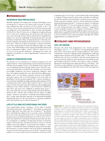
and CBX7) have never been validated as myeloma-driver genes. 15,16 Marrow stromal cell dependence
These same SNPs have also been independently associated with devel-
opment of MG, with the risk increasing with the number of alleles IL-6 dependence
carried. Moreover, the presence of hyperphosphorylated paratarg-7 Angiogenesis
17
(pP-7) carrier status has been reported as an autosomal dominantly Bone destruction
inherited risk factor for MG and myeloma in a European population
and in MG/myeloma cases in African Americans. 18,19 Migration and invasion
Increased proliferation index
LIFE STYLE AND OCCUPATIONAL FACTORS
Some epidemiologic studies, including a cohort study of Swedish Figure 107–1. Myeloma stages, from essential monoclonal gammo-
pathy (MG) to plasma cell leukemia. Myeloma evolves from a benign
and Finnish twins and a meta-analysis, 20,21 have shown an association condition called essential monoclonal gammopathy (or monoclonal
between high body mass index and risk of myeloma. 22,23 Specifically, gammopathy of undetermined significance), with an annual rate of
obese individuals have higher levels of cytokines, such as interleukin progression of 1 percent. In some patients, a stage called smoldering
(IL)-6 and insulin-like growth factor (IGF-1), which also are produced myeloma is sometimes evident, where there is a higher number of
by adipocytes and are potent growth factor for myeloma cells. No con- monoclonal plasma cells in the marrow, but still absence of symptoms.
24
sistent associations have been observed with any particular diet, alco- At early stages during the so-called intramedullary phase, myeloma
hol consumption, or smoking. Occupational exposure to pesticides, cells are totally dependent on marrow microenvironment to survive and
25
organic solvents (benzene, petroleum derivatives, styrene) or chronic on interleukin (IL)-6 and other cytokines. During progression, myeloma
radiation have been alleged to be associated with myeloma in some cells can acquire the capability of growing without microenvironmental
support and localize to other tissues (extramedullary disease) or circu-
studies, 25,26 but refuted by others. Furthermore, the use of thorium diox- late in the blood (secondary plasma cell leukemia). Active myeloma is
ide (thorotrast), a contrast medium used in the 1950s for angiography, characterized by onset of angiogenesis and bone lytic lesions in contrast
increases the risk of plasmacytomas up to fourfold. Exposure to acute to MG or smoldering myeloma; during late stages there is an increase
27
radiation, as in atomic bomb survivors increases the overall rate of MG in migration and invasion capabilities, as well as high proliferative rates.
Kaushansky_chapter 107_p1733-1772.indd 1734 9/21/15 12:33 PM