Page 1764 - Williams Hematology ( PDFDrive )
P. 1764

1738  Part XI:  Malignant Lymphoid Diseases                                         Chapter 107:  Myeloma            1739


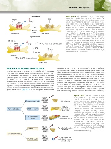
                                            Normal bone                          Figure 107–4.  Mechanism of bone remodeling in nor-
                                                                                 mal conditions and in the presence of myeloma cells. The
                                                              DKK1
                                                                                 major factors affecting osteoclast and osteoblast activa-
                             RANK         OPG                                    tion, and thus the balance between bone formation and
                                                                                 bone reabsorption, are illustrated in the upper panel.
                                                                                 Receptor activator of nuclear factor-κB (RANK) receptor/
                                              OPG                                receptor activator of nuclear factor-κB ligand (RANKL) and
                                                                                 macrophage inflammatory protein (MIP)-1α stimulate
                                                                                 osteoclastogenesis and osteoclast activity, while osteopro-
                                             RANKL
                       Osteoclasts                     Osteoblasts               tegerin (OPG) acts a decoy receptor for RANKL, reducing
                                                                                 its action. DKK1 (Dickkopf-1) is an inhibitor of osteoblast
                                           Myeloma bone                          activity. In the presence of myeloma cells in the bone, the
                                                                                 normal balance between osteoblasts and osteoclasts is
                                                                                 totally inverted. Specifically, myeloma cells secrete factors
                                                                                 to promote osteoclast activation, a result of upregulation
                          RANK          OPG         RANKL, DKK1, IL-6, ephrinB2/EphB4  of RANKL and MIP-1α, and to inhibit osteoblasts. Increased
                                                                                 levels of DKK1, activin, FRP-2 (frizzled related protein-2),
                                                OPG                              and sclerostin are evident in myeloma patients. In red are
                                                                DKK1             marked cytokines or receptors used as targets to treat mye-
                                                                                 loma bone disease. IL, interleukin.
                                                RANKL
                     Osteoclasts
                             Activin, FRP-2, sclerostin

                                                     Osteoblasts


                  PRECLINICAL MODELS OF MYELOMA                         subcutaneous injection of tumor myeloma cells in severe combined
                  Novel  therapies  need to  be  tested  in  preclinical  in vitro/vivo  models   immune deficiency (SCID) or nonobese diabetic (NOD)/SCID mice,
                                                                        which are immunocompromised. These models lack the complex mar-
                  capable of mimicking the role of human marrow microenvironment.   row–myeloma  interaction,  but  can  still  be used  to  explore  myeloma
                  In in vitro settings, myeloma cells are cocultured in liquid or semisolid   homing and novel drugs. Conversely, the SCID-hu or the SCID-rab
                  systems together with different cytokines (IL-6, IGF-1, TNF-α) or with   mice models recreate a look-alike microenvironment, able to sustain
                  autologous BMSCs from patients. However, these systems do not truly   myeloma cell growth.  Specifically, primary myeloma cells or mye-
                                                                                        224
                  recapitulate the marrow microenvironment 220–222  and  in vivo models   loma cell lines are grown in human fetal bone (the SCID-hu model)
                  are necessary. Two main types of myeloma animal models have been   or rabbit bone (SCID-rab model), later implanted in SCID mice.
                  exploited to study human myeloma biology and response to treatment:   Myeloma cells grow inside the implanted bones, or disseminate to
                  xenogeneic models in immunodeficient and humanized mice or syn-  the outer surface of the implanted bone, if they derive from patients
                                           223
                  geneic tumor models (Fig. 107–5).  The xenogeneic models require
                                                                        with  extramedullary  disease.  Moreover,  these  mice  have circulating
                                                                                           Figure  107–5.  Preclinical models of mye-
                                                                                           loma. Xenogeneic and syngeneic models
                                                                                           have been developed to study myeloma
                                      Subcutaneous model                    MM cells only  biology and test novel therapies. In xeno-
                                                                                           geneic models, human myeloma (MM) cells
                                                                                           are injected subcutaneously, inside human/
                                                                              MM cells +   rabbit fetal bone or into synthetic scaffolds,
                   Xenograft models     SCID-hu model                        Human/rabbit  previously coated with mesenchymal stromal
                                                                              fetal bone   cells (MSCs). In the last two types of models,
                                                                                           a look-alike microenvironment, able to sustain
                                                                                           myeloma cell growth, is present. Two types of
                                                                                           syngeneic models have been established: the
                                         SCID-synth-hu                        MM cells +
                                            model                          scaffold and MSCs  first one consists in the transplant of murine
                                                                                           myeloma cells into other mice (5TMM),
                                                                                           while the second one (Vκ*MYC mouse) is a
                                                                                           genetically  engineered mouse  where MYC
                                                                                           is activated in germinal center B-cells via an
                                                                                           activation-induced cytidine deaminase (AID)-
                                                                         Transplantable murine
                                         5TMM model                                        dependent  mechanism. SCID, severe com-
                                                                              MM cells
                                                                                           bined immune deficiency.
                   Syngeneic models
                                                                          AID-dependent MYC
                                         Vk*MYC model                     activation in germinal
                                                                            center B-cells.


          Kaushansky_chapter 107_p1733-1772.indd   1739                                                                 9/21/15   12:34 PM
   1759   1760   1761   1762   1763   1764   1765   1766   1767   1768   1769