
Page 1760 - Williams Hematology ( PDFDrive )
P. 1760
1734 Part XI: Malignant Lymphoid Diseases Chapter 107: Myeloma 1735
[t(6;14), t(11;14), and t(14;16)]. However, chromosome 13 deletions,
RAS mutations and non–immunoglobulin (Ig)-locus associated MYC
translocations are more frequent in myeloma. Indeed, the develop-
43
ment of myeloma seems to necessitate an immortalizing event, such as a
primary IGH translocation, an oncogene activation, or deregulation of Extramedullary myeloma
a tumor suppressor, to occur in the germinal center during the switch
recombination or somatic hypermutation, resulting in uncontrolled
42
expansion of a long-lived plasmablast/plasma cell. In early stages, Smoldering
myeloma cells are dependent on the growth support provided by bone MG myeloma Myeloma Plasma cell leukemia
marrow stromal cells (BMSCs) (intramedullary phase), but can become Hyperdiploidy
independent of their medullary environment at late stages (plasma cell
leukemia). However, 15 to 70 percent of newly diagnosed myeloma IGH translocations: t(11;14); t(4;14); MAF translocations
patients, using conventional morphology techniques 45–51 or multipara- Del (13q) and monosomy 13
52
metric flow cytometry have circulating clonotypic myeloma cells (cir-
culating tumor cells [CTCs]) in their blood, suggesting the presence of a chr (1q) amplification
“metastatic”/dissemination process that disseminates the disease hema- RAS mutations and myc overexpression
53
togenously. Moreover, the presence of CTCs in MG is a risk factor for
myeloma progression, 54,55 as well as a poor prognostic factor in newly Del (17p) or TP53 mutations
diagnosed or relapsed/refractory myeloma patients. 56,57 Myeloma CTCs RB1 mutations
share a similar phenotype to marrow myeloma cells, but are more quies-
cent, have better in vitro clonogenic capacity and have lower expression PTEN loss
of integrin and adhesion molecules (including CD138) making them p14 promoter meth
less dependent on marrow niches. 58
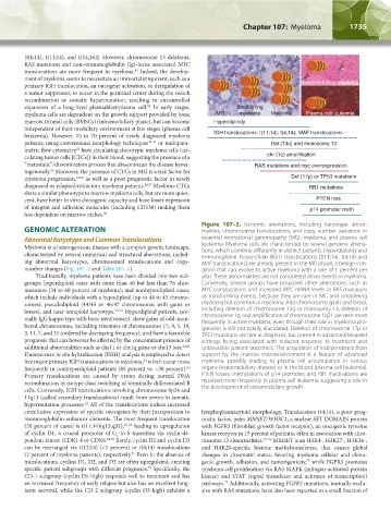
Figure 107–2. Genomic aberrations, including karyotype abnor-
GENOMIC ALTERATION malities, chromosomal translocations, and copy number variations in
Abnormal Karyotype and Common Translocations essential monoclonal gammopathy (MG), myeloma, and plasma cell
Myeloma is a heterogeneous disease with a complex genetic landscape, leukemia. Myeloma cells are characterized by several genomic aberra-
characterized by several numerical and structural aberrations, includ- tions, which combine differently in distinct patients. Hyperdiploidy and
immunoglobin heavy-chain (IGH) translocations [t(11;14), t(4;14) and
ing abnormal karyotypes, chromosomal translocations and copy- MAF translocations] are already present in the MG phase, a benign con-
number changes (Fig. 107–2 and Table 107–1). dition that can evolve to active myeloma with a rate of 1 percent per
Traditionally, myeloma patients have been divided into two sub- year. These abnormalities are not considered driver events in myeloma.
groups: hyperdiploid cases with more than 46 but less than 76 chro- Conversely, several groups have proposed other aberrancies, such as
mosomes (34 to 60 percent of myeloma); and nonhyperdiploid cases, MYC translocations and increased MYC mRNA levels or RAS mutations
which include individuals with a hypodiploid (up to 44 to 45 chromo- as transforming events, because they are rare in MG and smoldering
somes), pseudodiploid (44/45 or 46/47 chromosomes with gains or myeloma but common in myeloma. Also chromosome gains and losses,
losses), and near-tetraploid karyotype. 59–61 Hyperdiploid patients, nor- including deletion of chromosome 13q or monosomy 13, deletion of
mally IgG kappa-type with bone involvement, show gains of odd-num- chromosome 1p, and amplification of chromosome 1q21 are seen more
bered chromosomes, including trisomies of chromosomes 15, 9, 5, 19, frequently in active myeloma, even though their role in myeloma pro-
gression is still not totally elucidated. Deletion of chromosome 17p or
3, 11, 7, and 21 (ordered by decreasing frequency), and have a favorable TP53 mutations are rare at diagnosis, but present in advanced/relapsed
prognosis that can however be affected by the concomitant presence of settings, being associated with reduced response to treatment and
additional abnormalities such as chr11 or chr1q gains or chr13 loss. 62,63 unfavorable patient outcomes. The acquisition of independence from
Fluorescence in situ hybridization (FISH) analysis is employed to detect support by the marrow microenvironment is a feature of advanced
five major primary IGH translocations in myeloma, which occur more myeloma, possibly leading to plasma cell accumulation in various
64
frequently in nonhyperdiploid patients (85 percent vs. <30 percent). organs (extramedullary disease) or in the blood (plasma cell leukemia).
65
Primary translocations are caused by errors during normal DNA PTEN losses, methylations of p14 promoter, and RB1 inactivations are
recombination in isotype class switching of terminally differentiated B reported more frequently in plasma cell leukemia, suggesting a role in
cells. Conversely, IGH translocations involving chromosome 8p24 and the development of extramedullary growth.
11q13 (called secondary translocations) result from errors in somatic
hypermutation processes. All of the translocations induce increased
42
constitutive expression of specific oncogenes by their juxtaposition to lymphoplasmacytoid morphology. Translocation t(4;14), a poor prog-
immunoglobulin enhancer elements. The most frequent translocation nostic factor, pairs MMSET/WHSC1, a nuclear SET DOMAIN protein
(20 percent of cases) is t(11;14)(q13;q32), 66–68 leading to upregulation with FGFR3 (fibroblast growth factor receptor), an oncogenic tyrosine
of cyclin D1, a crucial promoter of G -to-S transition via cyclin-de- kinase receptor in 15 percent of patients, often in association with chro-
1
pendent kinase (CDK)-4 or CDK6. 69,70 Rarely, cyclin D2 and cyclin D3 mosome 13 abnormalities. 73–76 MMSET is an H3K4-, H3K27-, H3K36-,
can be rearranged via t(12;14) (<1 percent) or t(6;14) translocations and H4K20-specific histone methyltransferase, that causes global
(2 percent of myeloma patients), respectively. Even in the absence of changes in chromatin status, favoring myeloma cellular and clono-
71
translocations, cyclins D1, D2, and D3 are often upregulated, creating genic growth, adhesion, and tumorigenicity, while FGFR3 promotes
75
specific patient subgroups with different prognoses. Specifically, the myeloma cell proliferation via RAS-MAPK (mitogen-activated protein
72
CD-1 subgroup (cyclin D1-high) responds well to treatment and has kinase) and STAT (signal transducer and activator of transcription)
an increased frequency of early relapse but also has an excellent long- pathways. Additionally, activating FGFR3 mutations, mutually exclu-
77
term survival, while the CD-2 subgroup (cyclin D3-high) exhibits a sive with RAS mutations, have also been reported in a small fraction of
Kaushansky_chapter 107_p1733-1772.indd 1735 9/21/15 12:33 PM