
Page 200 - Williams Hematology ( PDFDrive )
P. 200
174 Part IV: Molecular and Cellular Hematology Chapter 13: Cytogenetics and Genetic Abnormalities 175
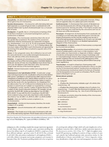
TABLE 13–1. Glossary of Cytogenetic Terminology
Aneuploidy—An abnormal chromosome number because of side of the centromere, it is called a paracentric inversion. If they
either gain or loss of chromosomes. were on opposite sides, it is called a pericentric inversion.
Banded chromosomes—Chromosomes with alternating dark and Isochromosome—A chromosome that consists of identical cop-
light segments as a result of special stains or pretreatment with ies of one chromosome arm with loss of the other arm. Thus, an
enzymes before staining. Each chromosome pair has a unique pat- isochromosome for the long arm of No. 17 [i(17)(q10] contains two
tern of bands. copies of the long arm (separated by the centromere) with loss of
Breakpoint—A specific site on a chromosome containing a DNA the short arm of the chromosome.
break that is involved in a structural rearrangement, such as a Karyotype—Arrangement of chromosomes from a particular cell
translocation or deletion. according to an internationally established system such that the
Centromere—The chromosome constriction that is the site of largest chromosomes are first and the smallest ones are last. A
the spindle fiber attachment. The position of the centromere normal female karyotype is described as 46, XX and a normal male
determines whether chromosomes are metacentric (X-shaped, karyotype is 46, XY. An idiogram is an idealized representation
e.g., chromosomes 1–3, 6–12, X, 16, 19, 20) or acrocentric (inverted (diagram) of the chromosomes.
V-shaped, e.g., chromosomes 13–15, 21, 22, Y). During mitosis, the Pseudodiploid—A diploid number of chromosomes accompanied
two exact copies of the DNA in each chromosome are separated by by structural abnormalities.
shortening of the spindle fibers attached to opposite sides of the Recurring Abnormality—A numerical or structural abnormality
dividing cell. noted in multiple patients who have a similar neoplasm. Such
Clone—In the cytogenetic sense, this is defined as two cells with abnormalities are characteristic or diagnostic of distinct subtypes
the same additional or structurally rearranged chromosome, or of leukemia and lymphoma that have unique morphologic and/
three cells with loss of the same chromosome. or immunophenotypic features. Recurring abnormalities represent
Deletion—A segment of a chromosome is missing as the result of genetic mutations that are involved in the pathogenesis of the
two breaks and loss of the intervening piece (interstitial deletion). corresponding diseases; many recurring abnormalities have prog-
Molecular studies of many recurring deletions have shown that, in nostic significance.
each case, the deletions were interstitial, rather than terminal Translocation—A break in at least two chromosomes with
(single break with loss of the terminal segment). exchange of material. In a reciprocal translocation, there is no obvi-
Diploid—Normal chromosome number and composition of ous loss of chromosomal material. Translocations are indicated by
chromosomes. t; the chromosomes involved are noted in the first set of brackets
Fluorescence in situ hybridization (FISH)—A molecular-cytoge- and the breakpoints in the second set of brackets. The Ph translo-
cation is t(9;22)(q34.1;q11.2).
netic technique based on the visualization of fluorescently-labeled
DNA probes hybridized to complementary DNA sequences from Nomenclature symbols:
metaphase or interphase cells, used to detect numerical and struc- p—Short arm
tural abnormalities. A short nomenclature description is used to q—Long arm
describe the results of in situ hybridization. For interphase FISH,
the abbreviation “nuc ish” is followed immediately by the locus des- +—If before the chromosome, indicates a gain of a whole chro-
ignation in parentheses (or multiple loci separated by a comma), mosome (e.g., +8)
a multiplication symbol, and the number of signals observed. The −—If before the chromosome, indicates a loss of a whole chro-
number of cells scored is placed in brackets. For example, normal mosome (e.g., −7) and if after the chromosome indicates loss of
results for the BCR-ABL1 probe are described as “nuc ish (ABL1, part of the chromosome (e.g., 5q−, loss of part of the long arm of
BCR)×2[400].” A case with the t(9;22) resulting in the BCR-ABL1 chromosome 5)
fusion analyzed using a dual-color, dual-fusion probe would be ?—Indicates uncertainty about the identity of the chromosome
described as “nuc ish (ABL1×3), (BCR×3), (ABL1 con BCR×2)[400].” or band listed just after the ?
Haploid—Only one-half the normal complement, i.e., t—translocation
23 chromosomes. del—deletion
Hyperdiploid—Additional chromosomes; therefore, the modal inv—inversion
number is 47 or greater.
Hypodiploid—Loss of chromosomes with a modal number of i—isochromosome
45 or less. mar—marker chromosome
Inversion—Two breaks occur in the same chromosome with rota- r—ring chromosome
tion of the intervening segment. If both breaks were on the same
Data from Rowley JD: Chromosome abnormalities in human cancer. In De Vita VT, Hellman S, Rosenberg S (eds): Practice and Principles of Oncol-
ogy 3rd ed. Philadelphia, PA: Lippincott Williams & Wilkins; 1991.
malignancies, often revealing complex but unique expression signatures of a novel genetic subtype of high-risk B-cell acute lymphocytic or lym-
for each disease subtype. For example, gene expression profiling has phoblastic leukemia (ALL), which has a similar expression signature as
shown that diffuse large B-cell lymphoma (DLBCL) comprises at least Ph-chromosome-positive ALL (Chap. 91). High-throughput array-
9,10
three different subtypes—germinal center B-cell (GCB)-like, activated based SNP genotyping, enabling the analysis of large numbers of cases
B-cell (ABC)-like, and primary mediastinal B-cell lymphoma (PMBL)— and controls, has facilitated genome-wide association studies for the
each with a distinct oncogenic mechanism, prognosis, and response to identification of disease susceptibility loci. Future diagnostic evalua-
11
therapies (Chap. 98). Expression profiling studies led to the recognition tion, identification of high-risk cases and management decisions will be
8
Kaushansky_chapter 13_p0173-0190.indd 175 17/09/15 6:32 pm