
Page 2189 - Williams Hematology ( PDFDrive )
P. 2189
2164 Part XII: Hemostasis and Thrombosis Chapter 126: von Willebrand Disease 2165
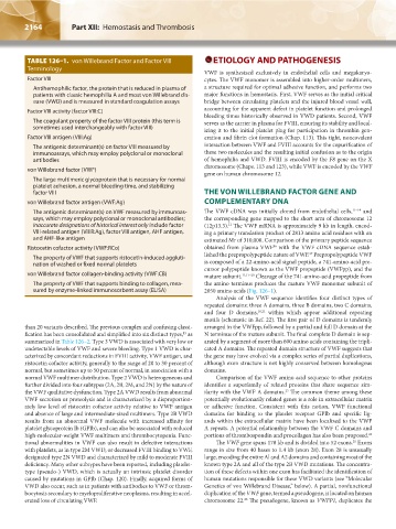
TABLE 126–1. von Willebrand Factor and Factor VIII ETIOLOGY AND PATHOGENESIS
Terminology
VWF is synthesized exclusively in endothelial cells and megakaryo-
Factor VIII cytes. The VWF monomer is assembled into higher-order multimers,
Antihemophilic factor, the protein that is reduced in plasma of a structure required for optimal adhesive function, and performs two
patients with classic hemophilia A and most von Willebrand dis- major functions in hemostasis. First, VWF serves as the initial critical
ease (VWD) and is measured in standard coagulation assays bridge between circulating platelets and the injured blood vessel wall,
Factor VIII activity (factor VIII:C) accounting for the apparent defect in platelet function and prolonged
bleeding times historically observed in VWD patients. Second, VWF
The coagulant property of the factor VIII protein (this term is serves as the carrier in plasma for FVIII, ensuring its stability and local-
sometimes used interchangeably with factor VIII)
izing it to the initial platelet plug for participation in thrombin gen-
Factor VIII antigen (VIII:Ag) eration and fibrin clot formation (Chap. 113). This tight, noncovalent
The antigenic determinant(s) on factor VIII measured by interaction between VWF and FVIII accounts for the copurification of
immunoassays, which may employ polyclonal or monoclonal these two molecules and the resulting initial confusion as to the origin
antibodies of hemophilia and VWD. FVIII is encoded by the F8 gene on the X
chromosome (Chaps. 113 and 123), while VWF is encoded by the VWF
von Willebrand factor (VWF)
gene on human chromosome 12.
The large multimeric glycoprotein that is necessary for normal
platelet adhesion, a normal bleeding time, and stabilizing
factor VIII THE VON WILLEBRAND FACTOR GENE AND
von Willebrand factor antigen (VWF:Ag) COMPLEMENTARY DNA
The antigenic determinant(s) on VWF measured by immunoas- The VWF cDNA was initially cloned from endothelial cells, 11–14 and
says, which may employ polyclonal or monoclonal antibodies; the corresponding gene mapped to the short arm of chromosome 12
inaccurate designations of historical interest only include factor (12p13.3). The VWF mRNA is approximately 9 kb in length, encod-
11
VIII-related antigen (VIIIR:Ag), factor VIII antigen, AHF antigen, ing a primary translation product of 2813 amino acid residues with an
and AHF-like antigen estimated Mr of 310,000. Comparison of the primary peptide sequence
16
Ristocetin cofactor activity (VWF:RCo) obtained from plasma VWF with the VWF cDNA sequence estab-
lished the prepropolypeptide nature of VWF. Prepropolypeptide VWF
17
The property of VWF that supports ristocetin-induced aggluti-
nation of washed or fixed normal platelets is composed of a 22-amino-acid signal peptide, a 741-amino-acid pre-
cursor polypeptide known as the VWF propeptide (VWFpp), and the
von Willebrand factor collagen-binding activity (VWF:CB) mature subunit. 11,17–20 Cleavage of the 741-amino-acid propeptide from
The property of VWF that supports binding to collagen, mea- the amino terminus produces the mature VWF monomer subunit of
sured by enzyme-linked immunosorbent assay (ELISA) 2050 amino acids (Fig. 126–1).
Analysis of the VWF sequence identifies four distinct types of
repeated domains: three A domains, three B domains, two C domains,
and four D domains, 18,21 within which appear additional repeating
motifs (schematic in Ref. 22). The first pair of D domains is tandemly
than 20 variants described. The previous complex and confusing classi- arranged in the VWFpp, followed by a partial and full D domain at the
fication has been consolidated and simplified into six distinct types, as N terminus of the mature subunit. The final complete D domain is sep-
15
summarized in Table 126–2. Type 3 VWD is associated with very low or arated by a segment of more than 600 amino acids containing the tripli-
undetectable levels of VWF and severe bleeding. Type 1 VWD is char- cated A domains. The repeated domain structure of VWF suggests that
acterized by concordant reductions in FVIII activity, VWF antigen, and the gene may have evolved via a complex series of partial duplications,
ristocetin cofactor activity, generally to the range of 20 to 30 percent of although exon structure is not highly conserved between homologous
normal, but sometimes up to 50 percent of normal, in association with a domains.
normal VWF multimer distribution. Type 2 VWD is heterogeneous and Comparison of the VWF amino acid sequence to other proteins
further divided into four subtypes (2A, 2B, 2M, and 2N) by the nature of identifies a superfamily of related proteins that share sequence sim-
the VWF qualitative dysfunction. Type 2A VWD results from abnormal ilarity with the VWF A domains. The common theme among these
23
VWF secretion or proteolysis and is characterized by a disproportion- potentially evolutionarily related genes is a role in extracellular matrix
ately low level of ristocetin cofactor activity relative to VWF antigen or adhesive function. Consistent with this notion, VWF functional
and absence of large and intermediate-sized multimers. Type 2B VWD domains for binding to the platelet receptor GPIb and specific lig-
results from an abnormal VWF molecule with increased affinity for ands within the extracellular matrix have been localized to the VWF
platelet glycoprotein Ib (GPIb), and can also be associated with reduced A repeats. A potential relationship between the VWF C domains and
high-molecular-weight VWF multimers and thrombocytopenia. Func- portions of thrombospondin and procollagen has also been proposed. 24
tional abnormalities in VWF can also result in defective interactions The VWF gene spans 178 kb and is divided into 52 exons. Exons
25
with platelets, as in type 2M VWD, or decreased FVIII binding to VWF, range in size from 40 bases to 1.4 kb (exon 28). Exon 28 is unusually
designated type 2N VWD and characterized by mild to moderate FVIII large, encoding the entire Al and A2 domains and containing most of the
deficiency. Many other subtypes have been reported, including platelet- known type 2A and all of the type 2B VWD mutations. The concentra-
type (pseudo-) VWD, which is actually an intrinsic platelet disorder tion of these defects within one exon has facilitated the identification of
caused by mutations in GPIb (Chap. 120). Finally, acquired forms of human mutations responsible for these VWD variants (see “Molecular
VWD also occur, such as in patients with antibodies to VWF or throm- Genetics of von Willebrand Disease,” below). A partial, nonfunctional
bocytosis secondary to myeloproliferative neoplasms, resulting in accel- duplication of the VWF gene, termed a pseudogene, is located on human
erated loss of circulating VWF. chromosome 22. The pseudogene, known as VWFP1, duplicates the
26
Kaushansky_chapter 126_p2163-2182.indd 2164 9/21/15 3:14 PM