
Page 2193 - Williams Hematology ( PDFDrive )
P. 2193
2168 Part XII: Hemostasis and Thrombosis Chapter 126: von Willebrand Disease 2169
113
colocalizes with VWF in the subendothelium. Type VI collagen sup- 2A
2A
ports the binding of VWF under high shear through cooperative inter- 2N 2M 2M
2M
actions between binding domains within the VWF A1 and A3 repeat. 2N 2A 2N 2M 2B2A 2M 2M
114
2A
2A
2A
Although VWF binding has also been demonstrated to a number of S D1 D2 D′ D3 A1 A2 A3 D4 B B B C1 C2 CI CK
1 2 3
other potential components of the subendothelium, including gly- VWF Exon 3–10 11–17 18–20 20–28 Exon 28 28–32 35–39 40–42 42–48 49–52
118
117
cosaminoglycans, 115,116 sulfatides, and VWF itself, the biologic
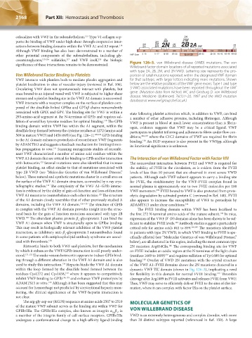
significance of these interactions remains to be demonstrated. Figure 126–3. von Willebrand disease (VWD) mutations. The von
Willebrand factor domain locations of all reported mutations associated
with type 2A, 2B, 2M, and 2N VWD. Lettering size represents the pro-
Von Willebrand Factor Binding to Platelets portion of total mutations reported within the designated VWF domain
VWF interacts with platelets both to mediate platelet aggregation and for that subtype, with larger letters indicating more mutations. Shown
platelet localization to sites of vascular injury (reviewed in Ref. 106). below are the relative positions of the VWF gene exons. Type 1 and type
Circulating VWF does not spontaneously interact with platelets, but 3 VWD associated mutations have been reported throughout the VWF
once bound to an injured vessel wall VWF is subjected to higher shear gene. (Mutation data from Nichols WC and Ginsburg D: von Willebrand
stresses and a platelet-binding site in the VWF A1 domain is uncovered. disease. Medicine (Baltimore) 76(1):1–20, 1997 and the VWD mutation
VWF interacts with a receptor complex on the surface of platelets com- database at www.vwf.group.shef.ac.uk.)
posed of the disulfide-linked GPIbα and GPIbβ chains noncovalently
associated with GPIX and GPV. The binding site for VWF is within a state following platelet activation which, in addition to VWF, can bind
293-amino-acid segment at the N-terminus of GPIb and requires sul- a number of other adhesive proteins, including fibrinogen. Although
119
fation of several key tyrosine residues for optimal binding. The GPIb VWF is present in blood at much lower concentrations than is fibrin-
binding domain within VWF lies within the A1 segment, within the ogen, evidence suggests that VWF may be a critical ligand. VWF
disulfide loop formed between the cysteine residues at 1272 (amino acid participates in platelet tethering and adhesion to fibrin under flow con-
509 in mature VWF) and 1458 (695) (see Fig. 126–1). 120,121 GPIb binding ditions, 104,137 where the C1C2 domains of VWF are required for fibrin
to the A1 domain enhances proteolysis of recombinant VWF fragments binding. An RGD sequence is also present in the VWFpp, although
138
by ADAMTS13 and suggests a feedback mechanism for limiting throm- its functional significance is unknown.
bus propagation in vivo. Scanning mutagenesis studies of recombi-
122
nant VWF characterized a number of amino acid residues within the
VWF A1 domain that are critical for binding to GPIb and for interaction The Interaction of von Willebrand Factor with Factor VIII
with botrocetin. Several mutations were also identified that increase The noncovalent interaction between FVIII and VWF is required for
123
platelet binding, an effect similar to that of mutations associated with the stability of FVIII in the circulation, as is evident from the FVIII
type 2B VWD (see “Molecular Genetics of von Willebrand Disease,” levels of less than 10 percent that are observed in most severe VWD
below). These natural and synthetic mutations cluster in a small area on patients. Although each VWF subunit appears to carry a binding site
the surface of the VWF A1 domain structure, as revealed by x-ray crys- for FVIII, the stoichiometry for the VWF–FVIII complex found in
124
tallographic studies. The complexity of the VWF A1–GPIb interac- normal plasma is approximately one to two FVIII molecules per 100
tion is evidenced by the ability of gain-of-function and loss-of-function VWF monomers. FVIII bound to VWF is also protected from prote-
139
125
VWF A1 mutants to counterbalance each other in mice. The structure olytic degradation by activated protein C (reviewed in Ref. 140). FVIII
of the A1 domain closely resembles that of other previously studied A also appears to increase the susceptibility of VWF to proteolysis by
domains, including the VWF A3 domain. 126–128 The structure of GPIb ADAMTS13 under shear conditions. 141
in complex with the VWF A1 domain provides insight into the struc- The FVIII binding domain within VWF has been localized to
tural basis for the gain of function mutations associated with type 2B the first 272 N-terminal amino acids of the mature subunit. In mice,
142
VWD. The abundant plasma protein β -glycoprotein I can bind the expression of the VWF D’-D3 domains alone has been shown to be suf-
129
2
VWF A1 domain when VWF is structurally open to GPIbα binding. ficient to stabilize FVIII levels. Antibody studies suggest a particularly
143
This may result in biologically relevant inhibition of the VWF-platelet critical role for amino acids 841 to 859. 144,145 The mutations identified
interaction, as inhibitory anti–β -glycoprotein I autoantibodies found in patients with type 2N VWD, in which VWF binding to FVIII is spe-
2
in some patients with antiphospholipid antibody syndrome are associ- cifically affected (see “Molecular Genetics of von Willebrand Disease,”
ated with thrombosis. 130 below), are all clustered in this region, including the most common type
Ristocetin binds to both VWF and platelets, but the mechanism 2N mutation Arg854Gln. The corresponding binding site for VWF
146
by which it enhances the VWF/GPIb interaction is still poorly under- on FVIII includes an acidic region at the N-terminus of the light chain
stood. 131,132 The snake venom botrocetin appears to induce GPIb bind- (residues 1669 to 1689) and requires sulfation of Tyr1680 for optimal
147
ing through a different alteration in the VWF A1 domain and is also binding. Overlay of VWD 2N mutations with the crystal structure
148
used to study this interaction. Heparin binds the VWF A1 domain of the VWF A1–FVIII domains shows the 2N mutations clustered in a
128
within the loop formed by the disulfide bond formed between the dynamic VWF TIL’ domain (shown in Fig. 126–3), implicating a need
residues Cys1272 and Cys1458, where it appears to competitively for flexibility in this domain for normal FVIII binding. Thrombin
133
149
inhibit VWF binding to GPIb 134,135 and enhance VWF proteolysis by cleavage after Arg1689 in FVIII activates and releases FVIII from VWF.
ADAMTS13 in vitro. Although it has been suggested that this may Thus, VWF may serve to efficiently deliver FVIII to the sites of clot for-
136
account for hemorrhage not predicted by conventional heparin mon- mation, where it can complex with factor IXa on the platelet surface.
itoring, the clinical significance of the VWF-heparin interaction is
not clear.
The arg-gly-asp-ser (RGDS) sequence at amino acids 2507 to 2510 MOLECULAR GENETICS OF
of the mature VWF subunit serves as the binding site within VWF for
GPIIb/IIIa. The GPIIb/IIIa complex, also known as integrin α β , is VON WILLEBRAND DISEASE
IIb 3
a member of the integrin family of cell surface receptors. GPIIb/IIIa VWD is an extremely heterogeneous and complex disorder, with more
undergoes a conformational change to a high-affinity ligand-binding than 20 distinct subtypes reported (referenced in Ref. 150). A large
Kaushansky_chapter 126_p2163-2182.indd 2168 9/21/15 3:14 PM