
Page 2194 - Williams Hematology ( PDFDrive )
P. 2194
2168 Part XII: Hemostasis and Thrombosis Chapter 126: von Willebrand Disease 2169
number of mutations within the VWF gene have been identified (see Subgroups within type 1 VWD have been proposed based on the rel-
Fig. 126–3). However, because of both the genetic complexity of VWD ative levels of VWF present in the plasma and platelet pools, 156–159 but
and the practical considerations of VWF gene sequencing in most clin- with the exception in some circumstances for an accelerated VWF
160
ical settings, a VWF gene mutation is not required for the diagnosis of clearance phenotype (unofficially termed VWF type 1C ), subtype
151
VWD. A list is maintained by a consortium of VWD investigators distinctions in type 1 VWD are not generally used in clinical practice.
and can be accessed through the Internet at http://www.vwf.group.shef Type 1 VWF was previously assumed to simply represent the het-
.ac.uk/. These findings form the basis for the simplified classification erozygous form of type 3 VWD. However, in a large Canadian study,
151
of VWD outlined in Table 126–2 and used throughout this chapter. 48 percent of heterozygous carriers of type 3 VWD gene mutations
Types 1 and 3 VWD are defined as pure quantitative deficiencies of carried a diagnosis of type 1 VWD, while the remainder were asymp-
161
VWF that are either partial (type 1) or complete (type 3). Type 2 VWD tomatic. Furthermore, in two large studies of type 1 VWD families,
is characterized by qualitative abnormalities of VWF structure and/or numerous putative VWF mutations were identified, but very few were
function. The quantity of VWF found in type 2 VWD may be normal, predictive of VWF null alleles. 31,162 Thus, some, but probably not all (see
but it is usually mildly to moderately decreased (see Table 126–2). section Clinical Features below), type 1 VWD is the result of defects
The diagnosis of VWD, particularly type 1 VWD, can be con- within the VWF gene. Studies of type 1 VWD mutations in patients,
founded by the incomplete penetrance of the disease and the wide range in vitro, and in animal models have characterized diverse mechanisms
163
of VWF levels in normal populations (see “Laboratory Features” below). underlying type 1 VWD, including decreased VWF production, reten-
Nonpathogenic variation can impact laboratory assays in vitro, as is the tion of VWF in the ER, 164,165 impaired VWF secretion, 165,166 and decreased
case with Asp1472His, which alters VWF-ristocetin interactions but VWF survival. 163,166,167 Mutations that give rise to defective VWF subunits
has no impact on hemostasis in vivo. Ethnicity should also be con- that interfere in a dominant negative way with the normal allele may be
152
sidered. European ancestries were overrepresented in the studies which particularly likely to cause symptomatic VWD in the heterozygote. For
168
informed laboratory cutoffs, while African Americans exhibit generally example, mutations at several cysteine residues in the VWF D3 domain
higher VWF and FVIII levels. Additionally, there are numerous VWF and in the VWFpp of patients with moderately severe type 1 VWD. VWF
gene “mutations” previously thought to be causative of VWD which are carrying one of these mutations is retained in the ER, where it is proposed
now known to be common in African Americans 153,154 who have normal to exert a dominant negative effect on VWF derived from the normal
153
VWF and FVIII levels, including the Asp1472His variant. 155 allele via heterodimerization and degradation. 169,170
To date, most mutation studies and genetic linkage analysis of
Type 1 von Willebrand Disease type 1 VWD have been consistent with defects within the VWF gene.
Type 1 VWD is the most common form, accounting for approximately Although no single mutation can explain the majority of type 1 VWD,
70 percent of patients with the disease. Type 1 VWD is generally common VWF founder mutations can occur within populations, such as
autosomal dominant in inheritance and is associated with coordinate Tyr1584Cys identified in 14 percent of Canadian type 1 VWD patients,
reductions in FVIII, ristocetin cofactor activity, and VWF antigen and possibly a similar proportion of patients in Europe. 162,171 The
with maintenance of the full complement of multimers (Fig. 126–4). Tyr1584Cys mutation is associated with decreased VWF survival, likely
a result of increased susceptibility to proteolysis by ADAMTS13. 172–175
These large multicenter studies of type 1 VWD families found candi-
date VWF mutations in 63 to 70 percent of families with type 1 VWD,
leaving 37 to 40 percent of type 1 VWD index cases without a putative
mutation in VWF. 162,171 Cases with VWF gene mutations tend to be more
severe and highly heritable, while cases without an identifiable VWF
mutation generally have higher VWF antigen (VWF:Ag) levels (>30
31
IU/dL). In a large, multicenter European study of 150 type 1 VWD
families, about one-third of cases historically diagnosed to have type
1 VWD were found to have abnormal multimers, and of these nearly
all (95 percent) had a putative VWF gene mutation and significantly
3 lower VWF:Ag, VWF ristocetin cofactor activity (VWF:RCo), assay of
FVIII activity (FVIII:C), and VWF collagen-binding assay (VWF:CB)
2 levels. Conversely, index cases with normal multimers had higher lab-
oratory VWF values and fewer identifiable VWF mutations (55 per-
cent), suggesting that the pathogenic mechanism(s) underlying this
1
162
cohort of “true” type 1 VWD patients is more genetically complex.
N 1 2A 2B 2A 2A 2A N Given the complex biosynthesis and processing of VWF, defects at a
(IIC) (IID) (IIE) number of other loci could also be expected to result in quantitative
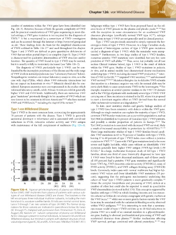
Figure 126–4. Agarose gel electrophoresis of plasma von Willebrand VWF abnormalities (reviewed in Ref. 176). This concept is supported by
factor (VWF). VWF multimers from plasma of patients with various sub- families with type 1 VWD in which bleeding histories and low ristoce-
types of von Willebrand disease (VWD) are shown. The brackets to the tin cofactor activities do not always cosegregate with genetic markers at
left encompass three individual multimer subunits, including the main the VWF locus, 31,177 while one or more genetic factors outside the VWF
band and its associate satellite bands. N indicates normal control lanes. locus may be associated with the variation in bleeding severity observed
Lanes 5 through 7 are rare variants of type 2A VWD. The former desig- within VWD pedigrees. 178,179 It is interesting to note that a spontane-
nations for these variants are indicated in parentheses below the lanes ous mouse model of type 1 VWD exhibits up to 20-fold reductions in
(IIC through E). (Adapted with permission from Zimmerman TS, Dent JA,
Ruggeri ZM, Nannini LH: Subunit composition of plasma von Willebrand plasma VWF as a result of an unusual mutation in a glycosyltransfer-
factor. Cleavage is present in normal individuals, increased in IIA and IIB von ase gene, leading to aberrant posttranslational processing of VWF and
180
Willebrand disease, but minimal in variants with aberrant structure of indi- accelerated clearance from plasma. Similar mechanisms affecting
vidual oligomers (types IIC, IID, and IIE). J Clin Invest 1986 Mar;77(3):947–951.) VWF survival, perhaps combined with altered proteolysis, 181–183 may
Kaushansky_chapter 126_p2163-2182.indd 2169 9/21/15 3:14 PM