
Page 220 - Williams Hematology ( PDFDrive )
P. 220
194 Part IV: Molecular and Cellular Hematology Chapter 14: Metabolism of Hematologic Neoplastic Cells 195
which is required for ribosome biogenesis. On the other hand, AMPK directly regulate genes involved in glycolysis, thereby linking an onco-
increases energy yield by stimulating glycolysis through phosphory- genic transcription factor to metabolism. Since these initial observa-
6,19
lation and activation of PFK-2. AMPK stimulates mitochondrial bio- tions, high-throughput methods have mapped MYC to a broad swath
genesis through phosphorylation of PGC1α and increases autophagy of metabolic enzyme genes involved in glycolysis, glutaminolysis, and
21
to recycle energy by phosphorylating ULK-1. Thus, increased AMPK lipogenesis. MYC also directly regulates genes involved in mitochon-
activity conserves energy and maximizes energy production. drial biogenesis and the production of ribosomes. Specifically, genes
Together with posttranscriptional responses to growth signal- highly induced by MYC include those involved in nucleolar function
ing and nutrients, the nuclear transcriptional response is necessary to and ribosome biogenesis, such as Ncl, NPM1, fibrillarin, and NOP52.
sustain the growth program through production of components of the Collectively, these studies uncover MYC’s role as a central regulator of
ribosome and mRNAs that give rise to all other components of the cell. cell growth through coupling of energy metabolism with cellular bio-
mTOR through its direct activation of specific transcription factors con- synthetic processes.
tributes to lipogenesis and mitochondrial biogenesis. Growth signaling Ribosome biogenesis is a critically important process for cell
also activates the MYC protooncogene, that regulates gene expression growth or cell mass accumulation. 25,26 Ribosomes are produced through
broadly to support cell growth and proliferation (see Figs. 14–2 and RNAs that are transcribed by RNA polymerases I (rRNA [ribosomal
19
14–3). Loss of function of Drosophila dMYC results in decreased cell RNA]), II (mRNA), and III (tRNAs [transfer RNAs] and small RNAs).
22
and body size, a phenotype that underscores MYC’s role in cell growth. rRNA is synthesized in the nucleolus from high copy numbers of rDNA,
This phenotype mimics the loss of ribosome protein gene function in a whose chromatin structure and transcription depends on nutrient
group of mutant flies termed Minutes. Hence, Drosophila genetics links availability. Under nutrient limitation, rDNA chromatin becomes less
26
MYC to cell growth control. Furthermore, MYC is the only transcrip- accessible, thereby restricting ribosome biogenesis. Ribosomal pro-
tion factor capable of stimulating the activity of RNA polymerases I, II, teins produced from mRNAs reenter the nucleolus, where components
and III, all of which are involved in ribosome biogenesis. of ribosomes are assembled into mature ribosomal particles, which are
MYC dimerizes with its partner Max to bind a specific DNA exported to the cytosol. The production of rRNAs and proteins also pro-
sequence, termed E-box (CACGTG), and activate transcription. It vides an opportunity for bioenergetic sensing of adequate nutrients to
23
can also inhibit transcription partly through direct binding to Miz-1 support nucleic acid and protein synthesis required for growth. In this
and diminishing the expression of Miz-1 target genes, including the regard, specific ribosomal protein subunits (RPL5, RPL11, and others)
cyclin-dependent kinase (CDK) inhibitor p21 and genes involved in can bind and inhibit MDM2 (mouse double minute 2 homolog), which
25
autophagy. Upon MYC activation, it is binding to proximal promoters binds to and mediates the degradation of p53. Thus, it is surmised that
accounts for most of it is transcriptional function in normal cells. When ribosomal proteins in excess of rRNAs would activate p53, triggering
MYC is experimentally expressed at levels comparable to those found checkpoints that block progression through the cell cycle, presumably
19
in cancers, excess MYC triggers p53-dependent checkpoints (see in response to nutrient limitation sensed as an imbalance in rRNA and
Fig. 14–3) that cause cell growth arrest or apoptosis. In multistep tum- ribosomal protein synthesis.
origenesis, therefore, p53 is often lost, unleashing MYC’s full oncogenic In addition to sensing ribosome biogenesis, p53 also responds to
potential. A high, unchecked level of MYC allows it to alter the tran- genotoxic stresses by directly regulating metabolism (see Fig. 14–2).
scriptome by amplifying selected target genes. MYC was first shown to P53, in general, activates oxidative phosphorylation and inhibits gly-
24
colysis. P53 can activate HK, which phosphorylates glucose in the
27
first step of glycolysis, and stimulate TIGAR that shunts glucose to
the pentose phosphate pathway through decreasing the levels of
fructose-2,6-bisphosphate (F2,6BP), which allosterically activates PFK.
P53 also increases the efficiency of mitochondrial function through
induction of cytochrome c oxidase (SCO ). Overall, it appears that the
28
2
normal function of p53 is to rewire metabolism to mitigate oxidative
Normal stress through increased production of NADPH and the antioxidant glu-
tathione. Gain of p53 function through specific mutations, on the other
hand, appears to alter metabolism through specific target genes that are
involved in cholesterol biosynthesis or phospholipase function. 29,30
Other tumor suppressors are also involved in metabolism (see Fig.
14–2). PTEN negatively modulates PI3K, and hence its loss stimulates
+MYC the PI3K pathway that is a potent regulator of cell metabolism through
+P53 P53 stimulation of glycolysis and activation of mTOR, AKT, MYC, and
31
HIF. The tumor suppressor LKB1, which is lost in some lung cancers,
21
Neoplastic normally activates the AMPK pathway and diminishes lipogenesis.
Loss of the von Hippel-Lindau (VHL) tumor suppressor activates HIF,
32
which transcriptionally regulates glucose metabolism. In addition to
any direct roles they play in regulating the cell-cycle machinery, tumor
suppressors—similar to protooncogenes—also regulate metabolism. By
coopting cellular responses to growth factor stimulation in the presence
of nutrients, activation of oncogenes and disablement of tumor sup-
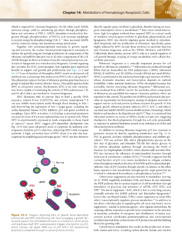
Figure 14–3. Diagram depicting fates of growth factor-stimulated
normal cells and MYC transforming cell. Upon engaging a growth fac- pressors achieve coordinated posttranscriptional and transcriptional
tor (pink square), the stimulated cell reacts as a bioreactor, which grows mobilizations that drive nutrients into ATP production and the building
and duplicates itself. MYC overexpression triggers checkpoints via p53, blocks for growing cells.
which induces cell death. With loss of p53 MYC’s full transforming Growth factor stimulation also results in the production of meta-
potential is unleashed through its transcriptional activities. bolic wastes and toxins, including carbon dioxide, lactate, and reactive
Kaushansky_chapter 14_p0191-0202.indd 195 17/09/15 6:35 pm