
Page 2247 - Williams Hematology ( PDFDrive )
P. 2247
2222 Part XII: Hemostasis and Thrombosis Chapter 130: Hereditary Thrombophilia 2223
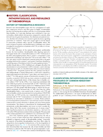
HISTORY, CLASSIFICATION, XIa
PATHOPHYSIOLOGY, AND PREVALENCE
OF THROMBOPHILIA
HISTORY OF THROMBOPHILIA RESEARCH
IXa + VIIIa APC + PS
Research into thrombophilia began with the investigation of candi-
date coagulation proteins and their genes in highly thrombophilic
families and linking abnormalities with the clinical phenotype within
these families. As a next step, findings were confirmed in case-con-
trol studies, which yielded risk increases compared to controls, often
derived from the general population. For clinicians and patients how- TF-VIIa Xa + Va Thrombin/IIa Fibrin
ever, absolute risk estimates were needed to guide decisions regarding
prevention or treatment. These were sought again in family studies
of consecutive probands with a specific thrombophilic defect. The
major progress in genetic and bioinformatics techniques now allows AT
investigation in populations of patients with VTE, as well as in throm-
bophilic families. 12–14 Figure 130–1. Regulation of blood coagulation. Coagulation is initi-
ated by a tissue factor (TF)–factor VIIa complex that can activate factor
In 1965, deficiency of the natural anticoagulant antithrombin IX or factor X. At high TF concentrations, factor X is activated primarily
became the first hereditary thrombophilia when Egeberg reported a by the TF-VIIa complex, whereas at low TF concentrations, the contri-
Norwegian family with a remarkable tendency to VTE. Deficiencies of bution of the factor IXa–factor VIIIa complex to the activation of factor
15
the other anticoagulant proteins, that is, protein C and protein S, were X becomes more pronounced. Coagulation is maintained through the
discovered as hereditary risk factors for VTE in the early 1980s. 16,17 By activation by thrombin of factor XI. The coagulation system is regulated
that time, genes could be cloned and numerous mutations in the genes by the protein C pathway. Thrombin activates protein C. Together with
encoding antithrombin, protein C, and protein S had been identified as protein S, activated protein C (APC) is capable of inactivating factors Va
underlying causes of low plasma levels of the anticoagulant proteins. 18–20 and VIIIa, which results in a downregulation of thrombin generation and
Another decade later, in 1993, Dahlbäck and colleagues described the consequently in an upregulation of the fibrinolytic system. The activity
phenomenon of activated protein C (APC) resistance, a poor anticoag- of thrombin is controlled by the inhibitor antithrombin. The solid arrows
indicate activation and the broken arrows inhibition.
ulant response to APC, in a Swedish family with an increased tendency
to develop VTE. The genetic basis for this APC resistance was discov-
21
ered independently in several laboratories in 1995 and is caused by a number of clinical studies provided reliable estimates of the relative and
single point mutation in the factor V gene which was termed factor V absolute risk for VTE.
Leiden. 22–25 In 1996, genetic analysis of prothrombin revealed a G-to-A
transition at position 20210 that was more common in patients with CLASSIFICATION, PATHOPHYSIOLOGY AND
VTE and a strong family history of this disease than in healthy con- PREVALENCE OF COMMON HEREDITARY
trols without VTE. In the 1970s, it was found that individuals with
26
non–O blood group have an increased risk of VTE. Individuals with THROMBOPHILIA
27
non–O blood group have higher levels of von Willebrand factor (VWF) Deficiencies of the Natural Anticoagulants Antithrombin,
and factor VIII than people with blood group O, which was the pre- Protein C, and Protein S
sumed mechanism of increased risk. In 1995, data from the Leiden Deficiencies of the natural anticoagulants antithrombin, protein C, and
Thrombophilia Study, a case-control study of patients with VTE and protein S, were among the first established hereditary thrombophilias.
matched healthy controls, demonstrated that increased factor VIII (FVIII) For antithrombin and protein C, two types of deficiencies are distin-
activity, but not VWF activity, was independently associated with an guished. In type I deficiency, levels of both antigen and activity are
increased risk of VTE. Homocysteine is an intermediary amino acid
28
formed by the conversion of methionine to cysteine. Homocystinuria
or severe hyperhomocysteinemia is a rare autosomal recessive disorder TABLE 130–1. Prevalence of Common Hereditary
characterized by severe elevations in plasma and urine homocysteine Thrombophilia
concentrations. This disease is characterized by developmental delay,
osteoporosis, ocular abnormalities, and severe occlusive vascular dis- Patients with
ease. About half of the vascular complications are of venous origin. General Population VTE
29
Mild hyperhomocysteinemia was therefore studied as a risk factor for Antithrombin, protein S, 1% 42–44 7% 41
VTE in the 1990s and homocysteine levels exceeding the 95th percentile or protein C deficiency
of the normal population were confirmed to be a risk factor for VTE. 30 Factor V Leiden Whites 4–7% 46,118 21% 22
Since then, numerous genetic variants that increase the risk of Nonwhites 0–1%
VTE to a more or lesser extent have been identified and are variably
included in diagnostic panels of thrombophilia testing. Essentially, Prothrombin G20210A Whites 2–3% 56,119 6%
31
the majority of hereditary thrombophilias exert their effect either by Nonwhites 0–1%
upregulation of procoagulant clotting factors, or by downregulation of Elevated FVIII:c levels 11% 28 25% 28
anticoagulant factors (Fig. 130–1). An overview of the common hered- 30 30
itary thrombophilias that increase the risk at least twofold, and their Mild 5% 10%
prevalence in patients with VTE and in the general population is pre- hyperhomocysteinemia
sented in Table 130–1. For these more common thrombophilias a large FVIII, factor VIII; VTE, venous thromboembolism.
Kaushansky_chapter 130_p2221-2232.indd 2222 9/21/15 4:33 PM