
Page 2248 - Williams Hematology ( PDFDrive )
P. 2248
2222 Part XII: Hemostasis and Thrombosis Chapter 130: Hereditary Thrombophilia 2223
reduced and in type II, antigen levels are normal, but one or more func- anticoagulants are rare (see Table 130–1). In cohorts of consecutive
tional defects in the molecule lead to a decreased activity. Type II antith- patients with VTE, the prevalence of a deficiency of one of the natu-
41
rombin deficiencies are further subdivided according to the site of the ral anticoagulants is below 10 percent. In the general population, the
defect in antithrombin. The defect is located in the thrombin binding prevalence of the deficiencies combined is approximately 1 percent. 42–44
domain (i.e., the reactive site) in type IIa deficiency, in the heparin bind-
ing domain in type IIb deficiency, and type IIc deficiency comprises a Factor V Leiden/Factor V G1691A
32
pleiotropic group of mutations. Interestingly, patients with type IIb In 1993, Dahlbäck and colleagues first described APC resistance in a
21
deficiency seem to have a significantly lower risk of VTE than other Swedish family with a high tendency of VTE. In the original paper,
32
types. Protein S circulates in two forms: free protein S (approximately Dahlbäck proposed that APC resistance was best explained by an
40 to 50 percent) which functions as a cofactor for APC, and protein S hereditary deficiency of a previously unrecognized cofactor to APC,
bound to complement component C4b-binding protein. In type I defi- after having ruled out several possible mechanisms, including deficien-
ciency, total and free antigen levels and activity are all reduced, in type cies of protein S and protein C, or linkage with polymorphisms in the
II deficiency, total and free antigen are normal, but activity is reduced, FVIII or VWF genes. He then showed that this "cofactor" was identical
and in type III deficiency, activity and free antigen are reduced, while to coagulation factor V. Soon thereafter, several laboratories indepen-
21
total antigen is low to normal. Type I and type III are probably pheno- dently from each other reported the underlying genetic defect, a single
typical variants of the same disease as family members with the same G-to-A substitution in the gene of factor V at nucleotide position 1691,
DNA mutation can present with either type I or type III deficiency. resulting in an amino acid change from Arginine (Arg) to Glutamine
33
Whether this classification into various subtypes is truly clinically rel- (Gln) at position 506, the first cleavage site of factor Va for APC 22–25
evant for any of the deficiencies of the natural anticoagulants is largely (Fig. 130–2). The mutation was named factor V Leiden after the city
unknown. Moreover, most laboratory panels now only test the activity in the Netherlands in which the group with the first publication was
22
of antithrombin, protein C, or protein S, and thereby do not distinguish located. The proteolytic inactivation of activated factor V (FVa) is
between different types of deficiencies. Homozygous antithrombin defi- approximately 10 times slower for Gln506-FVa compared with Arg506-
45
ciency is extremely rare and the only reported cases involve type IIb FVa, which explains the partial, but not full, resistance to APC. Factor
34
deficiencies. Homozygous type I deficiency has never been described V Leiden is the most common hereditary thrombophilia. In unselected
in humans and is believed to be incompatible with life. Complete consecutive patients with VTE, the prevalence is 20 to 25 percent. The
22
35
antithrombin deficiency in knockout mice leads to embryonic death. prevalence in the general population varies considerably between differ-
Homozygous protein C and protein S deficiencies are also very rare ent ethnic groups. Factor V Leiden is very rare among Asians and Afri-
and these are associated with neonatal purpura fulminans and massive cans but has a high prevalence (approximately 5 percent) among whites
thrombosis. 36,37 In a similar fashion, warfarin-induced skin necrosis has (see Table 130–1). Within Europe, the prevalence is higher in the
46
46
been described in patients with heterozygous protein C or S deficien- north than in the south. This implies a founder effect that suggests that
cies after initiation of vitamin K antagonists (VKAs). 38,39 The concept is the mutation occurred after the separation of non-Africans from Afri-
that after VKA initiation vitamin K–dependent protein C and S levels cans and after the divergence of whites and Asians. Studies using linkage
drop sooner than levels of factors II, IX, and X, thereby temporarily disequilibria between factor V Leiden and specific markers indicate that
causing a paradoxal procoagulant state. This is, however, a rare clin- the mutation occurred around 21,000 years ago. The high prevalence
40
47
48
ical complication, possibly resulting from concomitant treatment with of factor V Leiden suggests an evolutionary benefit. The presumed
(low-molecular-weight) heparin in the acute phase of VTE. Deficien- mechanism is reduced peripartum and menstrual blood loss in affected
cies can be caused by a large number of mutations, that are recorded in female carriers. 49,50 More recently, factor V Leiden was associated with
occasionally updated databases. 18–20 Overall, deficiencies of the natural increased sperm counts and a shorter conception time in affected male
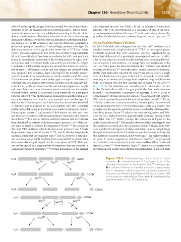
H N COOH Figure 130–2. Pathophysiology of the factor V Leiden
2
Arg 306 Arg 506 Arg 676 mutation. A. Activated protein C inactivates factor Va by
cleaving the protein at the Arginine (Arg) cleavage site.
506
B. In carriers of the factor V Leiden mutation, a point muta-
tion in the gene coding for factor V, causes replacement of
H 2 N COOH the amino acid Arginine by Glutamine (Gln) at position 506
of the protein, making factor Va resistant to inactivation by
Arg 306 Arg 979 activated protein C (i.e., APC resistance).
H 2 N COOH
H N COOH
2
Arg 306 Arg 679
Gln 506
H N COOH
2
Arg 306 Arg 679
N COOH
H 2
Kaushansky_chapter 130_p2221-2232.indd 2223 9/21/15 4:33 PM